

CAMBRIDGE, MA — In a discovery that defies long-standing principles of optics, researchers at the Massachusetts Institute of Technology (MIT) have harnessed “chaotic” laser light to create a powerful new imaging tool. Published in Nature Methods on April 27, 2026, the study reveals how high-power lasers can self-organize into a stable, ultra-fast “pencil beam.” This technology allows scientists to visualize the human blood-brain barrier (BBB) in 3D approximately 25 times faster than current industry standards, potentially shaving years off the development of treatments for Alzheimer’s, ALS, and brain tumors.
The research, led by Assistant Professor Sixian You and lead author Honghao Cao, represents a significant leap forward in “multiphoton imaging”—a technique used to peer deep into living tissue. By transforming what was once considered useless optical “noise” into a precision instrument, the team has provided a new window into one of the body’s most complex and protective structures.
Overcoming the “Chaotic” Barrier
For decades, a fundamental assumption in physics held that as you increase the power of a laser traveling through a multimode optical fiber, the light inevitably becomes chaotic. This “spatial-temporal instability” typically results in a scattered, blurry mess, making high-resolution imaging impossible at high speeds.
However, the MIT team, in collaboration with Harvard University and Beth Israel Deaconess Medical Center, discovered that under specific conditions, the system’s own physics undergoes a remarkable transition. Instead of scattering, the light pulses synchronized to form a single, tightly focused beam.
“The common belief in the field is that if you crank up the power in this type of laser, the light will inevitably become chaotic. But we proved that this is not the case,” says Sixian You, Assistant Professor in MIT’s Department of Electrical Engineering and Computer Science.
The researchers described this phenomenon as the system working in their favor. By utilizing this self-localized beam, they could scan biological samples with unprecedented efficiency without needing the bulky, expensive optical engineering typically required to stabilize high-power lasers.
Why the Blood-Brain Barrier Matters
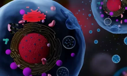
To understand why speed matters, one must understand the “fortress” it is trying to observe. The blood-brain barrier (BBB) is a highly selective layer of cells that protects the brain from toxins and pathogens. While vital for survival, it is a primary “gatekeeper” that blocks nearly 98% of small-molecule drugs and almost 100% of large-molecule therapies.
Current imaging methods used to study how drugs attempt to cross this barrier are often painstakingly slow. Researchers frequently have to take multiple 2D “slices” and stack them together to create a 3D image. This process is often too slow to capture the real-time biological “handshake” that occurs when a cell absorbs a drug.
The MIT “pencil beam” changes the math:
-
Speed: 25x faster than existing volumetric (3D) imaging.
-
Resolution: Maintains cellular-level detail, allowing researchers to see individual cells interacting with experimental therapies.
-
Real-Time Tracking: Enables scientists to watch how cells absorb drugs as it happens, rather than looking at “snapshots” after the fact.
Implications for Public Health and Drug Discovery
For the millions of families affected by neurological disorders, this discovery offers a new light at the end of the tunnel. While this is not a “cure” in itself, it is a critical piece of infrastructure for the laboratories that design cures.
In diseases like Alzheimer’s or brain cancer, the biggest hurdle is often not finding a drug that works in a petri dish, but finding one that can actually reach the brain in a living human. By speeding up the imaging process, researchers can more quickly discard drug candidates that fail to penetrate the BBB, allowing them to focus resources on the most promising therapies.
“This is a technical catalyst,” notes Dr. Elena Rossi, a neuro-imaging specialist not involved in the study. “If you can see the failure of a drug in days rather than months, you accelerate the entire pipeline of drug discovery. It moves the needle for public health by making the research process significantly more efficient.”
A Balanced Perspective: The Road Ahead
Despite the excitement, experts urge a measured perspective. This is currently a research tool, not a device found in a local hospital.
-
Preclinical Focus: The study demonstrated success in controlled laboratory environments and engineered tissue models.
-
Validation Needed: Transitioning this technology from a specialized MIT lab to a clinical setting where it could be used on human patients involves years of regulatory hurdles and broader validation across different types of imaging hardware.
-
Not a Diagnostic Tool (Yet): Currently, the “pencil beam” is designed to help scientists build better drugs, not to help doctors diagnose patients in a standard check-up.
Summary for the Health-Conscious Consumer
If you or a loved one is following the progress of neurological research, this study is a “behind-the-scenes” victory. It means the tools used to test the next generation of brain medicine just got a massive upgrade.
While it doesn’t change your current treatment plan or daily health habits, it represents a shift in how we understand the brain’s most stubborn defenses. By turning the “chaos” of laser light into a precision tool, science has found a faster way to see through the barriers that have long kept brain diseases so difficult to treat.
Medical Disclaimer: This article is for informational purposes only and should not be considered medical advice. Always consult with qualified healthcare professionals before making any health-related decisions or changes to your treatment plan. The information presented here is based on current research and expert opinions, which may evolve as new evidence emerges.
References
- https://www.earth.com/news/chaotic-laser-beam-turned-into-a-powerful-brain-imaging-tool/


