

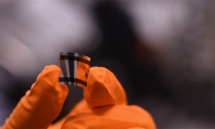
CHICAGO — In a development that shifts the conversation around artificial intelligence from clinical assistance to cybersecurity, a groundbreaking study published on March 24, 2026, reveals that synthetic X-ray images are now sophisticated enough to deceive both veteran radiologists and the world’s most advanced AI models. The research, appearing in the journal Radiology, warns that these “medical deepfakes” expose a critical vulnerability in the global healthcare infrastructure, potentially opening the door to insurance fraud, legal manipulation, and diagnostic sabotage.
A New Frontier of Deception
The study, led by Mickael Tordjman, M.D., a postdoctoral fellow at the Icahn School of Medicine at Mount Sinai, provides the most comprehensive look to date at how generative AI can fabricate medical reality. Researchers tested 264 chest X-ray images—a 50-50 split between authentic patient scans and synthetic images generated by tools including ChatGPT (GPT-4o) and RoentGen.
To evaluate the “believability” of these fakes, the team recruited 17 radiologists from 12 hospitals across six countries. The cohort represented a broad spectrum of expertise, from residents to specialists with 40 years of experience. In a blinded setup, the results were sobering:
-
Initial Detection: Only 41% of radiologists spontaneously identified the AI-generated images as unusual when tasked with assessing technical quality.
-
Prompted Accuracy: Even after being informed that synthetic images were present, the radiologists’ detection accuracy rose to only 75%.
-
Experience Gap: Perhaps most surprisingly, there was no significant difference in detection rates between novice doctors and those with decades of experience.
“These deepfake X-rays are realistic enough to deceive radiologists, the most highly trained medical image specialists, even when they were aware that AI-generated images were present,” Dr. Tordjman stated.
The fallibility extended to machines as well. Leading AI models, including GPT-5, Gemini 2.5 Pro, and Llama 4 Maverick, showed detection accuracies ranging from 57% to 85%. Paradoxically, even ChatGPT-4o struggled to identify the very images it had helped create.
The Anatomy of a Medical Fake
While the synthetic images were highly convincing, researchers noted that they often lacked the “messiness” of human biology. Dr. Tordjman pointed out several telltale signs that current AI models struggle to replicate:
-
Symmetry and Smoothness: Synthetic lungs often appeared too symmetrical, and bones appeared unnaturally smooth.
-
The “Perfect” Spine: AI tended to generate spines that were perfectly straight, failing to account for the natural curvature or minor scoliosis common in the general population.
-
Uniformity: Blood vessels in AI images often displayed a uniform distribution that ignores the biological variability found in actual patients.
However, as generative models evolve, these artifacts are expected to disappear. The study warns that chest X-rays are merely “the tip of the iceberg,” with the technology likely moving toward 3D modalities such as CT and MRI scans in the near future.
Implications for Public Health and Litigation
The rise of medical deepfakes introduces a “high-stakes vulnerability,” according to the research team. Beyond the clinical risk of a doctor treating a non-existent condition—or missing a real one—the implications for the legal and insurance sectors are profound.
“We face a significant risk for fraudulent litigation,” Dr. Tordjman warned. “A fabricated fracture could be indistinguishable from a real one in a personal injury case.”
Furthermore, cybersecurity experts express concern over “injection attacks.” In such a scenario, hackers could breach a hospital network and replace real patient images with synthetics. This could be used to alter a high-profile patient’s diagnosis or simply to sow chaos within a healthcare system. For patients, this could lead to an erosion of trust in the digital medical records that underpin modern telemedicine and insurance claims.
Balancing Innovation with Integrity
Generative AI is not inherently a “villain” in medical imaging. For years, researchers have used synthetic data to augment small datasets, helping to train diagnostic AI for rare diseases where real patient images are scarce. This “dual-use” nature of the technology—capable of both accelerating medical breakthroughs and facilitating deception—presents a complex challenge for regulators.
The Radiological Society of North America (RSNA) emphasizes that while AI aids efficiency in an era of radiologist shortages, it now requires a new layer of verification. Unlike video deepfakes, which are often betrayed by unnatural movements (the “uncanny valley”), X-rays are static and grayscale. This makes the anomalies subtler and far more difficult for the human eye to catch during a busy shift.
Limitations and Counter-Perspectives
While the study’s findings are alarming, some experts urge a measured response. Critics of the “panic” narrative note that in controlled settings, radiologists still perform better than random chance (50%).
“Currently, many AI outputs remain ‘too perfect’ for routine deception in a clinical environment where radiologists are looking for specific patient histories,” noted one independent reviewer.
Additionally, the study focused exclusively on chest X-rays. It remains to be seen if the same levels of deception can be maintained in more complex, multi-layered imaging like PET scans. However, the fact that experience did not improve detection suggests that “intuition” is not a defense against high-quality synthetics; only specific, technical training on AI artifacts seems to help.
Safeguarding the Future of Diagnostics
To combat this emerging threat, the research team and the RSNA have proposed several immediate safeguards:
-
Digital Watermarking: Embedding invisible, cryptographic signatures into images at the moment of capture.
-
Blockchain Provenance: Utilizing decentralized ledgers to track an image from the X-ray machine to the doctor’s workstation, ensuring it has not been tampered with in transit.
-
Specialized Training: Educating radiologists on the specific “AI signatures” identified in this study.
-
Forensic Algorithms: Deploying secondary AI tools, such as the DSKI model, specifically designed to detect synthetic patterns that the human eye might miss.
As AI becomes ubiquitous in the laboratory and the clinic, the medical community must now treat image integrity as a pillar of patient safety. The goal is to preserve the benefits of AI innovation while building a “digital immune system” to protect the sanctity of the medical record.
Medical Disclaimer
This article is for informational purposes only and should not be considered medical advice. Always consult with qualified healthcare professionals before making any health-related decisions or changes to your treatment plan. The information presented here is based on current research and expert opinions, which may evolve as new evidence emerges.
References
- https://www.reuters.com/business/healthcare-pharmaceuticals/health-rounds-fake-x-rays-created-by-ai-fool-radiologists-even-ai-itself-2026-03-25/


