
Published: February 9, 2026
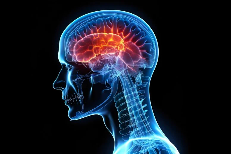
NEW YORK — In a breakthrough that bridges the gap between ancient survival instincts and modern artificial intelligence, researchers at NYU Langone Health have identified the specific brain region responsible for “one-shot learning”—the sudden flash of insight that allows us to recognize a blurry or distorted object instantly. The study, published this week in Nature Communications, pinpoints the high-level visual cortex (HLVC) as the seat of this perceptual “aha!” moment, offering new hope for understanding the mechanics of hallucinations in conditions like schizophrenia and Parkinson’s disease.
The Mystery of the “One-Shot”
Human survival has long depended on the ability to make sense of incomplete information. Whether it was an ancestor spotting a predator camouflaged in tall grass or a modern driver recognizing a pedestrian through a fogged windshield, the brain’s ability to “fill in the blanks” is a primal necessity.
Despite decades of study, the mechanism behind this—termed perceptual one-shot learning—has remained elusive. Unlike traditional learning, which requires repetition (like practicing a piano scale), one-shot learning happens in an instant. Once you “see” the hidden image, you cannot “un-see” it.
“Our work revealed not just where these ‘priors’—stored images from our past—are kept, but also the specific brain computations involved in accessing them,” says co-senior study author Biyu He, Ph.D., an associate professor at NYU Grossman School of Medicine.
Mapping the Mind’s Library
To track this mental phenomenon, the research team used Mooney images: high-contrast, black-and-white pictures that look like meaningless splotches until a clear version of the image is revealed. Once the participant sees the clear photo, their brain uses that “prior” information to decode the splotches instantly.
The researchers employed a multi-pronged approach to find where the brain stores these visual cheat sheets:
-
fMRI Imaging: Tracked blood flow to identify active brain cells.
-
Intracranial EEG (iEEG): Used electrodes (with the help of patients already undergoing neurosurgery) to capture rapid-fire signaling that fMRI might miss.
-
Behavioral Testing: Researchers rotated and resized images to see what information the “priors” actually contained.
The team discovered that while changing the size of an image didn’t hinder recognition, rotating it did. This suggested that our internal library of priors stores specific patterns and shapes rather than abstract concepts. The earliest neural signaling changes occurred in the HLVC, confirming it as the gateway where stored memories meet incoming visual data.
When Intuition Goes Awry: Hallucinations
The implications of this discovery extend far beyond basic biology. In a healthy brain, there is a delicate balance between what our eyes see and what our “priors” tell us. However, in certain neurological disorders, this balance shifts.
“Past studies have shown that patients with schizophrenia and Parkinson’s disease have abnormal one-shot learning,” explains Dr. He. In these cases, the stored “priors” can become too dominant, overwhelming the actual sensory input from the eyes. This leads the brain to “see” things that aren’t there—the foundation of a hallucination.
By identifying the HLVC as the hub for this process, researchers now have a “directly testable theory” to investigate why the brain generates these false perceptions.
Bridging the Gap to Artificial Intelligence
The study didn’t just look at human brains; it also looked at silicon ones. The team built a Vision Transformer, an AI model designed to find patterns in images.
While AI has historically struggled with one-shot learning—usually requiring thousands of examples to recognize a cat—the researchers added a specific “prior” module to their model. This allowed the AI to store image information and use it to recognize new, distorted data, mimicking human performance more closely than any previous model.
“We now anticipate the development of AI models with human-like perceptual mechanisms,” says co-senior author Eric Oermann, M.D. “This is more evidence of a growing convergence between computational neuroscience and advances in AI.”
What This Means for You
While this research is rooted in complex neuroscience, its daily applications are profound. Understanding how the brain uses “priors” can help us appreciate how our expectations shape our reality.
For the general public, this study underscores the importance of cognitive health. While we cannot “train” our HLVC directly like a muscle, staying mentally active and exposing ourselves to diverse visual stimuli helps build the library of “priors” that our brain relies on to navigate an unpredictable world.
“This is the beginning of understanding the ‘aha!’ moment,” says Dr. Sarah J. Morrison, a clinical neurologist not involved in the study. “If we can map how the brain makes sense of a blurry image, we are one step closer to understanding how we solve complex problems or even experience creative inspiration.”
Key Findings at a Glance
| Feature | Finding |
| Primary Brain Region | High-level Visual Cortex (HLVC) |
| Mechanism | Accessing “priors” (stored templates) to decode new data |
| Clinical Link | Overactive “priors” may explain hallucinations in schizophrenia |
| AI Impact | Leads to more efficient “one-shot” machine learning models |
References
-
Study Citation: Hachisuka, A., et al. (2026). Neural and computational mechanisms underlying one-shot perceptual learning in humans. Nature Communications. DOI: 10.1038/s41467-026-68711-x.
Medical Disclaimer: This article is for informational purposes only and should not be considered medical advice. Always consult with qualified healthcare professionals before making any health-related decisions or changes to your treatment plan. The information presented here is based on current research and expert opinions, which may evolve as new evidence emerges.


