
In a milestone for regenerative medicine that could fundamentally change the trajectory of neonatal surgery, researchers have successfully used lab-grown tissue to repair esophageal defects in large animal models. The study, published March 19, 2026, in Nature Biotechnology, demonstrates that personalized, bioengineered grafts can integrate into the body, restore swallowing function, and grow alongside the patient—all without the need for lifelong immunosuppressant drugs.
The breakthrough, led by teams at the UCL Great Ormond Street Institute of Child Health (UCL GOS ICH) and Great Ormond Street Hospital (GOSH), provides a potential “holy grail” for treating Long-Gap Esophageal Atresia (LGOA). This rare condition leaves newborns unable to swallow, often necessitating years of invasive surgeries and reliance on feeding tubes. With a 100% survival rate in the 30-day safety trial, researchers are now aiming for human clinical trials within the next five years.
The Challenge of the “Missing Link”
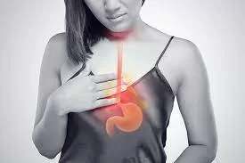
Esophageal atresia is a congenital condition where the esophagus—the muscular tube connecting the mouth to the stomach—fails to form completely during pregnancy. In about 10% of cases, known as “long-gap” atresia, the distance between the two ends of the pipe is too great to be sewn together directly.
Currently, surgeons must resort to “interposition” surgeries, which involve pulling the stomach up into the chest or using a segment of the patient’s colon to bridge the gap. While life-saving, these procedures are fraught with long-term complications, including:
-
Severe acid reflux that can damage the new lining.
-
Respiratory distress due to the repositioning of internal organs.
-
Aspiration pneumonia and chronic feeding difficulties.
-
Increased cancer risk in the transposed tissue later in life.
For families like those of two-year-old Casey McIntyre of London, the current reality is a grueling marathon of medical interventions. Born with an 11 cm gap, Casey has undergone multiple procedures and is only now, years later, beginning the difficult process of learning how to swallow.
A Personalized Biological Solution
The new research moves away from “repurposing” other organs and instead focuses on building a functional esophagus from scratch. The process begins with a “scaffold”—a donor pig esophagus that has been chemically stripped of all its original cells (decellularized). What remains is a natural, tubular matrix of collagen that provides the perfect structural blueprint for human anatomy.
To prevent the body from rejecting the graft, researchers use the patient’s own cells to “re-seed” the scaffold:
-
Biopsy: A small sample of muscle and skin-like cells (fibroblasts) is taken from the patient.
-
Expansion: These cells are grown in a laboratory for several weeks until billions are available.
-
Maturation: The cells are placed onto the scaffold inside a bioreactor—a high-tech chamber that mimics the conditions of the human body, providing nutrients and “exercise” to the developing tissue.
“After successful implantation, our grafts grew, matured, and began to function like native tissue,” says Dr. Natalie Durkin, pediatric surgical registrar at GOSH and the study’s lead author. “Each one of these steps represents a key milestone in being able to deliver this as a viable treatment option for children in the near future.”
Proving Function: The Minipig Study
The researchers tested the grafts in eight minipigs, chosen because their esophageal size and physiology closely resemble those of human infants. The results were remarkably consistent. All animals survived the initial 30-day safety period and were able to begin oral feeding on the very first day after surgery.
Over a six-month follow-up period, the bioengineered tubes did more than just hold steady; they transformed. High-tech imaging and spatial transcriptomics revealed that the grafts had developed organized layers of muscle, nerves, and blood vessels. Crucially, the grafts exhibited peristalsis—the wave-like muscle contractions necessary to push food down to the stomach—averaging 7.2 seconds per bolus.
“Our technology could allow us to build a child a new esophagus using their own cells,” explains Dr. Marco Pellegrini, co-leader of the study. This “autologous” approach eliminates the need for the toxic immunosuppression drugs usually required after transplants.
Expert Insights and Caveats
While the results are being hailed as a “game-changer,” independent experts urge a balance of optimism and scientific caution.
Dr. Jennifer Smith, a pediatric gastroenterologist at Boston Children’s Hospital who was not involved in the study, noted that avoiding donor-recipient mismatches is a massive win for neonates with immature immune systems. However, she points out that human healing is often more complex than in animal models.
“The study showed that several animals developed strictures (narrowing of the tube) that required balloon dilation,” Dr. Smith observed. “While this is common in human infants as well, we must ensure these lab-grown tissues can withstand the decades of wear and tear a human child will put them through.”
Dr. Alan Fine, a regenerative medicine specialist at Nationwide Children’s Hospital, highlighted the scalability of the research. “Pig scaffolds are already used safely in heart valves. If we can standardize the production of these grafts for different infant sizes, this could eventually become an ‘off-the-shelf’ solution for surgeons.”
The Road to 2031: What This Means for Patients
For healthcare professionals, this shift from “palliative bridging” to “regenerative repair” could significantly reduce the cost and duration of neonatal intensive care. For parents, it offers the hope of a “one-and-done” surgery that allows their child to eat, grow, and play like any other toddler.
“The idea that there could be one operation early on would be life-changing,” shared Casey McIntyre’s parents, reflecting on the potential for future families to avoid the years of feeding tubes they have endured.
Professor Paolo De Coppi, NIHR Nuffield Professor of Paediatric Surgery at UCL GOS ICH, remains confident in the timeline. “With the success of this research, we hope that we can be successfully offering an engineered tissue alternative to children who desperately need it within five years.”
The researchers are now focusing on automating the production of these grafts and developing ways to track the cells more precisely after implantation to ensure long-term safety.
Reference Section
Primary Study:
-
Durkin N, et al. “Autologous tissue-engineered oesophageal graft for repair of long-gap oesophageal atresia.” Nature Biotechnology. Published March 19, 2026. DOI: 10.1038/s41587-026-00936-8.
Medical Disclaimer: This article is for informational purposes only and should not be considered medical advice. Always consult with qualified healthcare professionals before making any health-related decisions or changes to your treatment plan. The information presented here is based on current research and expert opinions, which may evolve as new evidence emerges.


