
NEW DELHI — In a major breakthrough for medical diagnostics, scientists in India have unveiled a novel light-based imaging platform that functions like a biological “chalkboard,” allowing researchers to map an unprecedented number of proteins within a single tissue sample.
Announced by the Press Information Bureau (PIB) on May 26, 2026, the technology—named Cleavable Light-Erased Antibody Reporter (CLEAR)—overcomes a decades-old bottleneck in medical microscopy. Developed by a research team at the Jawaharlal Nehru Centre for Advanced Scientific Research (JNCASR) in Bengaluru, an autonomous institute of the Department of Science and Technology (DST), this method enables deep, high-resolution visual tracking of the complex molecular networks driving aggressive cancers and debilitating neurobiological conditions.
The Molecular Map Dilemma
To understand human disease, pathologists must look closely at proteins—the primary orchestrators of cellular function. From signaling a tumor to grow to triggering the degradation of brain cells, proteins serve as both the diagnostic markers of a disease and the ultimate targets for therapeutic drugs.
Historically, doctors trying to diagnose complex diseases from patient tissue biopsy samples faced a rigid physical boundary known as the “spectral limit.” Conventional immunofluorescence imaging relies on attaching glowing fluorescent dyes (fluorophores) to antibodies, which then bind to specific target proteins. Because different dyes emit distinct colors under a microscope, scientists can typically look at only three to five proteins at a time before the color spectra overlap, resulting in a blurry, unreadable visual mess.
For modern precision medicine, looking at five proteins is no longer enough. Pathologists require a comprehensive spatial map showing exactly where hundreds of different proteins are located relative to one another within their native environments.
How CLEAR Turns Light into an Eraser
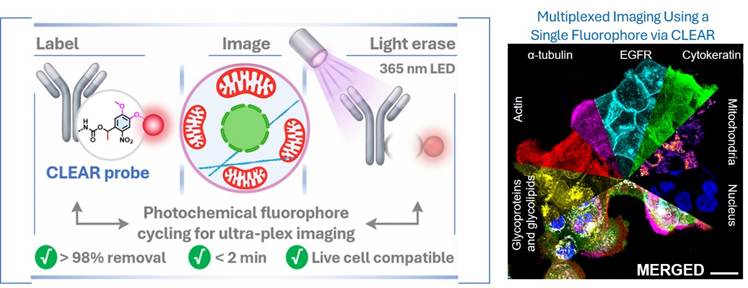
The JNCASR team, led by principal investigator Prof. Sarit S. Agasti, resolved this limitation by designing a method that reuses the exact same color window over and over again. Instead of trying to pack dozens of different color dyes into a delicate live cell, CLEAR uses a single fluorescent marker attached to a light-cleavable probe system.
The technology relies on an elegant, iterative loop that functions similarly to writing, erasing, and rewriting on a blackboard:
-
Step 1: Label & Image: Scientists introduce CLEAR probes to a sample, labeling a specific set of target proteins with fluorescent tags, and capturing high-resolution images under a microscope.
-
Step 2: Light Cleanse: The sample is exposed to a gentle, non-toxic pulse of 365-nanometer (nm) LED ultraviolet light. This precise wave of light safely snaps the chemical bond holding the fluorescent marker to the antibody.
-
Step 3: Wash Away: The cleared fluorescent signal is washed out cleanly, removing more than 98% of the fluorescence in under two minutes.
-
Step 4: Repeat: The structural framework of the cell remains perfectly intact, allowing scientists to instantly introduce a new round of antibody probes targeting a completely different set of proteins in the exact same sample window.
[Label Proteins] ──> [Image under Microscope] ──> [365 nm LED Pulse (Erase)] ──> [Wash & Repeat]
By continuously repeating this cycle, pathologists can stack images on top of one another to build a highly detailed, multi-layered molecular map.
Transforming Oncology and Neurobiology
“This approach fundamentally redefines multiplexed imaging by enabling virtually unlimited protein visualization without requiring multiple fluorophores,” noted the JNCASR research group in their study, published in the peer-reviewed journal Chemical Science (Kalita et al., 2026).
The clinical implications for early disease detection and tailored healthcare are profound:
1. Cracking the Cancer Microenvironment
Tumors are highly complex, rapidly mutating ecosystems composed of malignant cells, structural tissue, and invading immune cells. Standard biopsies often fail to capture this diversity. By utilizing CLEAR to map dozens of cellular markers simultaneously, pathologists can identify the specific sub-type of a patient’s cancer, predict how aggressively it will spread, and determine if it will respond to advanced therapies like immunotherapy.
2. Tracing Neurodegenerative Breakdown
In neurological diseases like Alzheimer’s or Parkinson’s, specific toxic protein aggregates disrupt vast networks of delicate synapses (the junctions where brain cells communicate). Because CLEAR is uniquely gentle and entirely compatible with delicate biological samples—including live cells—it allows neuroscientists to watch real-time structural degradation and organelle shifts without destroying the fragile brain tissue sections being studied.
3. Deconstructing Immune Responses
In collaboration with researchers at the Indian Institute of Science (IISc), Prof. Agasti’s team successfully validated the CLEAR platform within complex, dynamic immune cell systems. The researchers demonstrated high-dimensional interrogation of coordinated cellular restructuring during the formation of an “immunological synapse”—the critical event where an immune cell recognizes a foreign threat.
Overcoming the Blind Spots of Existing Technology
While multi-protein imaging is an expanding field, older techniques carry steep tradeoffs. Some current methods use harsh chemical stripping agents or heavy-metal isotopes to erase or identify signals, which frequently degrades the sample tissue, distorts the natural spatial distribution of proteins, and limits the number of cycles a scientist can execute.
Other cutting-edge solutions rely on complex DNA-barcoded antibody structures. While highly effective, these DNA-based systems require intricate synthetic engineering, strict sequence design, and highly delicate temperature or chemical washing controls, making them operationally difficult to implement at scale in a standard diagnostic hospital lab.
CLEAR stands out because it uses conventionally conjugated antibodies and handles signal clearing remotely using a mild light source. This drastically reduces processing times, eliminates the need for complex fluid-handling infrastructure, and minimizes sample degradation.
Current Limitations and the Path to the Clinic
Despite its immense promise, independent medical experts emphasize that several hurdles must be cleared before the technology leaves the research bench for local hospital pathology departments.
A primary challenge is speed and throughput automation. Performing multiple iterative cycles manually can be labor-intensive. For CLEAR to become a standard tool for everyday clinical diagnosis, the process must be integrated into fully automated fluidics and microscope imaging systems that can run overnight without human intervention. Furthermore, the long-term chemical storage stability of the specialized light-cleavable probes must be evaluated to ensure they can be easily distributed to clinics worldwide.
Additionally, while a 365 nm LED light pulse is considerably gentler than harsh chemical stripping, scientists will need to rigorously confirm that repeated light exposure does not alter rare, low-abundance biological markers across a variety of human tissue types during exceptionally long imaging cycles.
References
-
Ministry of Science & Technology, Government of India. (2026, May 26). “CLEAR” technology can revolutionize protein imaging & facilitate detection of Cancer and Neurobiological Diseases. Press Information Bureau (PIB), Delhi. https://www.pib.gov.in/PressReleasePage.aspx?PRID=2265421
Medical Disclaimer: This article is for informational purposes only and should not be considered medical advice. Always consult with qualified healthcare professionals before making any health-related decisions or changes to your treatment plan. The information presented here is based on current research and expert opinions, which may evolve as new evidence emerges.


