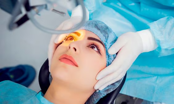
Uveitis, a rare inflammatory disease affecting the choroid of the eye, poses significant challenges for diagnosis and monitoring due to its diverse subtypes and variable presentation. However, a recent study conducted by researchers from the University Hospital Bonn, in collaboration with experts from Berlin, Münster, and Mannheim, sheds light on a promising imaging technique that could revolutionize the management of posterior uveitis and panuveitis.
Published in the journal Biomolecules, the study focuses on the application of fundus autofluorescence (FAF) as a diagnostic and monitoring tool for these challenging eye conditions. Dr. Maximilian Wintergerst from the Eye Clinic at the University Hospital Bonn explains, “Depending on the inflamed anatomical structure, this disease can be divided into the subtypes anterior, intermediate, posterior, and panuveitis. The exact diagnosis of posterior uveitis and panuveitis can be challenging, as there are many different and sometimes extremely rare subtypes.”
FAF, a non-invasive imaging technique, involves stimulating fluorophores in the eye tissue with a defined wavelength of light, resulting in the emission of fluorescence. This emission pattern provides valuable insights into the underlying pathology of uveitis, aiding in accurate diagnosis and assessment of disease activity. Dr. Matthias Mauschitz, Head of the Uveitis Clinic at the UKB, elaborates, “In unclear cases, this can help to make the correct diagnosis. In addition, the autofluorescence signal can also provide us with information on the current state of inflammation in certain forms of uveitis.”
The researchers emphasize the importance of choosing the appropriate wavelength for FAF imaging, as different wavelengths can yield varying results based on lesion depth and location. By combining multiple wavelengths, clinicians can obtain a comprehensive understanding of the disease process and tailor treatment accordingly.
“In some specific subtypes of uveitis, [FAF] can also provide important indications of a flare-up of inflammatory activity,” summarizes Wintergerst. The study underscores the pivotal role of FAF in enhancing the diagnostic accuracy and therapeutic management of posterior uveitis and panuveitis, while also highlighting avenues for future research, such as exploring the utility of multi-wavelength FAF imaging.
As efforts continue to unravel the complexities of uveitis, innovations in imaging technologies like FAF offer renewed hope for improved outcomes and quality of life for patients battling this debilitating eye condition.