
April 24, 2026
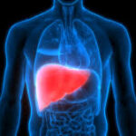
ROURKELA, INDIA – Researchers at the National Institute of Technology (NIT) Rourkela have announced the development of a patented bio-ink that could significantly improve the precision and biological success of 3D-bioprinted tissues. Reported in April 2026, the innovation addresses a persistent “tug-of-war” in regenerative medicine: creating a material that is sturdy enough to hold its shape after printing while remaining hospitable enough for living cells to thrive. While early laboratory results show particular promise for bone and cartilage repair, experts caution that the technology must navigate a rigorous multi-year path of animal and clinical trials before reaching hospital operating rooms.
The “Living Ink” Breakthrough
At the heart of the discovery is a high shape-fidelity, protein-polysaccharide composite. Developed by a team led by Prof. Devendra Verma in the Department of Biotechnology and Medical Engineering, the bio-ink utilizes a sophisticated blend of Bovine Serum Albumin, Sodium Alginate, and polyelectrolyte complexes of gelatin and chitosan.
In 3D bioprinting, the “ink” is not just a pigment but a structural scaffold that houses live cells. Historically, scientists have struggled with a trade-off: high-viscosity materials provide great structural “fidelity” (the ability to hold a shape) but often suffocate or crush cells. Conversely, thinner, more cell-friendly materials often collapse into a “puddle” before they can solidify.
“Our goal was to bridge the gap between printability and biological performance,” stated Prof. Verma in a recent release. By integrating protein-polysaccharide interactions with nanofibrous complexes, the team has created a system that flows smoothly through the printer nozzle but stabilizes almost instantly upon deposition.
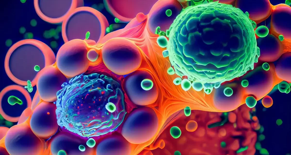
Promising Lab Signals: Mimicking Nature
The NIT Rourkela team’s findings, published in the International Journal of Biological Macromolecules, highlight the ink’s ability to mimic the extracellular matrix (ECM)—the natural “scaffolding” that surrounds cells in the human body.
Key Laboratory Findings:
-
High Cell Viability: Scaffolds tested in the lab demonstrated over 90% cell viability, meaning the vast majority of cells survived the high-pressure printing process.
-
Structural Integrity: The material maintained its 3D geometry without the need for harsh chemical “cross-linkers” that can often damage sensitive biological tissue.
-
Tissue Potential: Early tests showed the ink supported cell adhesion, proliferation, and even the synthesis of collagen—the primary structural protein in skin and bone.
The Long Road to the Clinic
Despite the excitement surrounding the patent (Patent No. 583759), the medical community remains pragmatically cautious. The transition from a successful “bench” experiment to a “bedside” treatment is a notorious hurdle in biotechnology.
“3D bioprinting is transformative, but achieving full vascularization—the growth of blood vessels within the tissue—remains a primary challenge,” noted researchers in a 2025 review in Biofabrication. Without a blood supply, larger printed tissues cannot receive oxygen or nutrients, leading to cell death in the center of the construct.
Furthermore, the NIT Rourkela bio-ink must still prove it can integrate seamlessly with a host’s immune system. While the materials used (like chitosan and alginate) are generally biocompatible, the long-term mechanical stability under the weight-bearing stress of a human knee or hip is yet to be tested in living subjects.
Public Health and Personalized Care
If the technology successfully clears animal and human trials, the implications for public health could be profound. Currently, patients with severe bone fractures or degenerative cartilage issues often rely on autografts (taking bone from another part of their body) or allografts (donor tissue). Both methods carry risks of infection, donor site pain, or tissue rejection.
A reliable, printable bio-ink would allow surgeons to:
-
Scan a patient’s specific injury using MRI or CT imaging.
-
Model a perfectly fitting replacement scaffold.
-
Print the repair tissue using the patient’s own cells, virtually eliminating the risk of rejection.
Beyond surgery, these bio-inks are being used to create “organ-on-a-chip” models. These are miniature, printed versions of human organs used to test new drugs, potentially reducing the pharmaceutical industry’s reliance on animal testing and speeding up the delivery of safe medications to the public.
What This Means for You
For the average consumer, it is important to view this as a significant “building block” rather than an immediate cure. While you cannot yet walk into a clinic and request a 3D-printed meniscus, the development at NIT Rourkela represents the hardening of the scientific foundation required to make that a reality in the next decade.
The next phase for the Rourkela researchers involves moving into animal models to observe how the bio-ink interacts with a complex, living immune system—a critical step before human volunteers are ever involved.
Reference Section
Peer-Reviewed Studies:
- https://health.economictimes.indiatimes.com/news/industry/nitr-team-develops-bio-ink-to-aid-3d-bioprinting-tissue-engineering/130454615?utm_source=top_story&utm_medium=homepage
Medical Disclaimer: This article is for informational purposes only and should not be considered medical advice. Always consult with qualified healthcare professionals before making any health-related decisions or changes to your treatment plan. The information presented here is based on current research and expert opinions, which may evolve as new evidence emerges.


