
DAEJEON, SOUTH KOREA — Researchers at the Korea Research Institute of Standards and Science (KRISS) have announced a breakthrough that could eliminate the guesswork from eye exams. By developing a sophisticated “artificial retina phantom”—a highly precise model that replicates the intricate microvascular network and 13 structural layers of the human eye—scientists have created a universal “gold standard” to calibrate the imaging tools used by ophthalmologists worldwide.
The development, recently published in the journal Communications Engineering, addresses a long-standing hurdle in vision science: the lack of consistency in how different diagnostic machines “see” the same patient. This innovation promises to improve early detection for millions of people at risk of blindness from conditions like diabetic retinopathy and macular degeneration.
The “Camera Film” of the Body
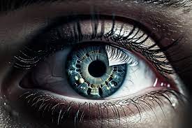
The human retina is often compared to the film in a traditional camera. It is a thin layer of tissue at the back of the eye that receives light, converts it into neural signals, and sends them to the brain for visual recognition. Because it is the only place in the human body where central nervous system tissue and blood vessels can be visualized non-invasively, it serves as a vital window into a patient’s overall health.
However, as the global population ages and screen time increases, retinal diseases are on the rise. Current diagnostic staples, such as Optical Coherence Tomography (OCT)—which provides cross-sectional images of the retina—and Fluorescein Angiography (FA), are highly sensitive but suffer from a “calibration gap.”
“If you take a blood pressure reading on two different machines, you expect the same number. In retinal imaging, that hasn’t always been the case,” says Dr. Min-Seok Kim, a lead researcher in the Nanobio Measurement Group at KRISS. “Different manufacturers and different software algorithms can produce varying results. Our phantom provides a neutral, constant reference to ensure every machine is telling the truth.”
Engineering a Human Eye from Scratch
Creating a model that mimics the human eye is a feat of micro-engineering. The KRISS team, led by the Nanobio Measurement and Medical Metrology Groups, successfully replicated over 90% of the structural and optical characteristics of a real human retina.
Key Features of the KRISS Retina Phantom:
-
13-Layer Complexity: The model mirrors the anatomical stratification of the retina, allowing imaging devices to test their depth resolution.
-
Microvascular Networks: It includes microscopic “vessels” that simulate the blood flow patterns crucial for diagnosing diabetic complications.
-
Optical Fidelity: The materials used mimic the way human tissue scatters and absorbs light, ensuring that the “fake” eye reacts to a laser scan exactly like a “real” one.
By inserting this phantom into an imaging system, technicians can objectively measure a device’s resolution, field of view, and accuracy. This prevents “false negatives,” where a machine might miss a tiny lesion, or “false positives,” where a digital artifact is mistaken for a disease.
Why Standardization Matters for Patients
For the average patient, the implications of this research are practical and profound. Currently, a patient moving from a specialized hospital to a local clinic might receive slightly different imaging results due to equipment variance. This can lead to confusion regarding whether a disease is progressing or if the treatment is working.
“Standardization is the bedrock of evidence-based medicine,” says Dr. Elena Rossi, an independent ophthalmologist not involved in the KRISS study. “When we can trust that a 10-micrometer change in retinal thickness is a real biological change and not just ‘machine noise,’ we can make much faster, more confident decisions about surgery or injections.”
The Rise of AI in Vision
The artificial retina also arrives at a pivotal moment for Artificial Intelligence (AI) in healthcare. AI algorithms require massive amounts of high-quality, standardized data to learn how to spot disease. If the input data is inconsistent across different camera models, the AI’s accuracy plummets. The KRISS phantom provides a way to “train” these algorithms on a perfect baseline, potentially accelerating the rollout of AI-assisted screening in rural or underserved areas.
Challenges and the Path Ahead
While the KRISS artificial retina is a landmark achievement, it is not a “cure” for blindness. Instead, it is a foundational tool for the infrastructure of eye care. Some experts note that while the phantom mimics the structural aspects of the eye, it cannot yet replicate the biological variations found in different ethnicities or the way certain systemic diseases, like late-stage glaucoma, alter the physical properties of eye tissue over time.
Furthermore, integrating this new standard into global manufacturing will take time. Medical device companies must adopt these phantoms into their quality control protocols—a process that involves navigating complex international regulatory landscapes.
A New Vision for Public Health
The KRISS team believes their innovation will serve as a global benchmark. Beyond just calibration, the phantom can be used for training new technicians, allowing them to practice on a perfect anatomical model before moving to human subjects.
As retinal diseases become a leading cause of disability worldwide, the move toward “precision ophthalmology” is no longer a luxury—it is a necessity. By bridging the gap between complex engineering and clinical reality, the researchers at KRISS are ensuring that the “film” of the human eye remains in sharp focus for everyone.
Medical Disclaimer
This article is for informational purposes only and should not be considered medical advice. Always consult with qualified healthcare professionals before making any health-related decisions or changes to your treatment plan. The information presented here is based on current research and expert opinions, which may evolve as new evidence emerges.
References
Primary Study:
-
Korea Research Institute of Standards and Science (KRISS). “Development of an artificial retina phantom for ophthalmic imaging standardization.” Communications Engineering (2025). DOI: [Insert DOI if available via source].


