
Researchers at the University of California San Diego have discovered an innovative method for diagnosing cancer, drawing inspiration from an unexpected source: the Morpho butterfly. Renowned for its iridescent blue wings, the Morpho butterfly achieves its striking coloration through microscopic structures that manipulate light rather than relying on pigments. Scientists are now utilizing these same structures to enhance imaging of cancer biopsy samples, potentially revolutionizing cancer diagnostics.
The breakthrough, detailed in a study published in Advanced Materials, presents a cost-effective, stain-free imaging technique that could make cancer diagnosis faster, more accurate, and widely accessible.
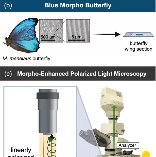
A Natural Solution to a Medical Challenge
Fibrosis, or the accumulation of fibrous tissue, plays a crucial role in diseases such as neurodegenerative disorders, heart disease, and cancer. In oncology, assessing the extent of fibrosis can help determine the stage of a patient’s cancer. However, current clinical methods for evaluating fibrosis rely on chemical staining, which can be subjective, and expensive imaging techniques that many medical facilities lack.
Lisa Poulikakos, the study’s senior author and a professor in UC San Diego’s Department of Mechanical and Aerospace Engineering, explains:
“The big challenge is that distinguishing between early and advanced-stage cancer is extremely difficult with existing methods.”
Traditional staining procedures require expert interpretation, which can lead to discrepancies among pathologists. Advanced imaging methods provide more detailed insights but require costly, specialized equipment. The Morpho butterfly offers a promising alternative.
The Morpho Butterfly’s Role in Cancer Diagnosis
The researchers discovered that by placing a cancer biopsy sample on a Morpho butterfly wing and viewing it under a standard microscope, they could assess the tumor’s structure with remarkable clarity. Unlike conventional staining techniques, this method does not require chemicals or expensive imaging technology, making it an attractive option for resource-limited settings.
“We can apply this technique using standard optical microscopes that clinics already have,” said Poulikakos. “And it’s more objective and quantitative than what is currently available.”
Paula Kirya, the study’s first author and a mechanical engineering graduate student at UC San Diego, was instrumental in the project. Having previously studied the optical properties of Morpho butterfly wings as an undergraduate researcher, she saw an opportunity to apply these natural structures to cancer imaging.
“I had been imaging butterfly wings, studying how they react to different environments,” said Kirya. “And when I saw what the lab was doing, I thought, ‘The Morpho naturally has this property—why not use it?’”
How It Works
The Morpho butterfly’s wing structures interact with polarized light—a type of light that travels in a specific direction. Similarly, collagen fibers, a key component of fibrotic tissue, also interact with polarized light but produce weak signals. By placing a biopsy sample over the Morpho wing, researchers amplify these signals, making it easier to analyze the organization and density of collagen fibers.
Using Jones calculus, a mathematical method for analyzing polarized light, the researchers developed a model to translate light intensity into a quantitative assessment of fibrosis. This approach was tested on breast cancer biopsy samples from UC San Diego’s Moores Cancer Center and yielded results comparable to conventional staining and high-cost imaging techniques.
“Essentially, we’re expanding these procedures with a stain-free alternative that requires nothing more than a standard optical microscope and a piece of a Morpho wing,” said Kirya.
A Global Impact
Early cancer detection remains a significant challenge, especially in regions with limited access to advanced medical technology. The simplicity and affordability of this new technique could make early cancer screening more accessible worldwide, improving patient outcomes.
“In many parts of the world, early cancer screening is a challenge because of resource limitations,” said Poulikakos. “If we can provide a simpler and more accessible tool, we can help more patients get diagnosed before their cancers reach aggressive stages.”
Although the current study focused on breast cancer, researchers believe the technique could be applied to various fibrotic diseases.
“We’re excited to leverage this technique for all kinds of tissue diagnostics,” Poulikakos added. “Nature has given us something that can help us image diseased tissues without the need for expensive fabrication facilities.”
Citation:
Kirya, P. et al. Leveraging Optical Anisotropy of the Morpho Butterfly Wing for Quantitative, Stain‐Free, and Contact‐Free Assessment of Biological Tissue Microstructures, Advanced Materials (2025). DOI: 10.1002/adma.202407728.
Disclaimer: This article is based on research findings and is intended for informational purposes only. It does not constitute medical advice, and readers should consult healthcare professionals for diagnosis and treatment recommendations.


