
Routine eye exams could soon become a frontline tool in detecting Alzheimer’s disease decades before cognitive symptoms arise, according to new research led by neuroscientists at The Jackson Laboratory and published in the journal Alzheimer’s & Dementia in July 2025. The researchers studied mice with a common genetic mutation and found subtle changes in the retina’s blood vessels may mirror processes in the brain that precede Alzheimer’s—raising hope for accessible, non-invasive early diagnosis.
Key Findings and Emerging Evidence
The study centers on mice carrying the MTHFR677C>T gene variant, which affects up to 40% of the human population. Investigators discovered that retinas in these mice showed twisted blood vessels, narrowed and swollen arteries, and reduced branching as early as six months of age—features that have been linked to problems with blood flow and increased dementia risk.
Proteomics and imaging revealed that molecular changes in the retina closely mirror alterations seen in the brain—supporting the hypothesis that one’s retina reflects the health status of central nervous system tissues. As lead researcher Alaina Reagan explained, “If you’re at an optometrist or ophthalmologist appointment, and they can see odd vascular changes in your retina, that could potentially represent something that is also happening in your brain…very informative for early diagnostics”.
Expert Commentary and Context
Dr. Muralidharan Sargurupremraj, an Alzheimer’s researcher at UT Health San Antonio (not involved in the study), notes, “The concept that vascular changes in the retina may serve as a surrogate marker for brain disease is compelling. Our work also supports a causal link between small vessel vascular injury and dementia—making non-invasive ophthalmic imaging potentially valuable for screening and prevention strategies”.
Routine retinal imaging could identify those at heightened genetic risk years before forgetfulness, personality changes, or language problems—the classic symptoms of Alzheimer’s—emerge. The retina’s accessibility via the pupil means practitioners can observe nervous tissue in a “window into the brain,” positioning eye exams as practical, affordable tools in neurology and geriatrics.
Public Health Implications
With dementia prevalence projected to reach 75 million globally by 2030, early detection is a public health imperative. If retinal blood vessel changes can reliably signal the disease process, routine eye exams could be integrated into preventive care, enabling lifestyle changes, timely interventions, and clinical trial participation for at-risk individuals.
For health-conscious individuals, the findings underscore the value of regular eye checkups, particularly for middle-aged adults with risk factors or family history of dementia. Promoting retinal imaging—ideally as part of annual optometry visits—may help flag those who would benefit from memory clinics or genetic counseling.
Limitations and Counterarguments
While the study’s results are promising, several caveats warrant attention. The primary data comes from mouse models rather than human clinical trials, and genetic risk does not guarantee disease development—many people with the MTHFR677C>T mutation never develop Alzheimer’s. Moreover, retinal changes may result from hypertension, diabetes, or other chronic conditions, complicating the use of this biomarker in isolation.
Some experts caution that more research is needed to validate these findings in large, diverse human populations and to determine which retinal features are most predictive of Alzheimer’s vs. other causes of cognitive decline. Robust, longitudinal studies are required before clinical guidelines will recommend retinal imaging as a routine screening tool for dementia.
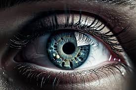
Understanding the Science: Retina as a Biomarker
The retina is part of the central nervous system and, like the brain, contains neurons and small blood vessels vulnerable to the same disease processes. Imaging techniques have evolved to detect thinning of retinal layers, microvascular anomalies, and protein deposits—all of which have shown associations with neurodegenerative disorders in research. Analogy: Just as a mechanic can spot engine problems by observing exhaust, doctors may one day assess brain health by looking into the eye.
Practical Takeaways for Readers
-
Schedule regular eye exams: Especially for adults over 40 or those with a family history of dementia.
-
Discuss findings with healthcare providers: If retinal vascular anomalies are found, further neurological assessment may be warranted.
-
Promote vascular health: Controlling blood pressure, cholesterol, and glucose levels benefits both eye and brain health, potentially lowering the risk of dementia.
Balanced Perspective
As the science advances, routine eye exams could add a valuable layer to dementia risk assessment. However, individuals should not rely solely on eye findings or self-diagnose. Continued dialogue between ophthalmologists, neurologists, and primary care clinicians is vital for translating research into practical preventive care.
Medical Disclaimer
Medical Disclaimer: This article is for informational purposes only and should not be considered medical advice. Always consult with qualified healthcare professionals before making any health-related decisions or changes to your treatment plan. The information presented here is based on current research and expert opinions, which may evolve as new evidence emerges.
Reference Section
- https://health.economictimes.indiatimes.com/news/industry/routine-eye-tests-may-reveal-early-alzheimers-signs-study-suggests/123546044?utm_source=top_story&utm_medium=homepage


