

January 2, 2026
LONDON — Researchers have achieved a landmark breakthrough in personalized medicine by developing the first “lung-on-chip” model composed entirely of genetically identical human cells. The study, led by the Francis Crick Institute in collaboration with biotechnology firm AlveoliX, successfully recreated the complex environment of the human lung’s air sacs using stem cells from a single donor. By simulating the rhythmic expansion and contraction of breathing, this “breathing” chip allows scientists to observe the early, hidden stages of infections like tuberculosis (TB) with unprecedented accuracy, potentially signaling a shift away from animal testing toward hyper-personalized treatment plans.
Recreating the Breath of Life
At the heart of the human respiratory system are the alveoli—tiny, delicate air sacs where oxygen enters the blood and carbon dioxide leaves it. These sacs are more than just gas exchangers; they are the primary battleground where the body fights off inhaled pathogens like influenza and TB.
For years, scientists have used “organ-on-chip” technology—microscopic channels etched into plastic to house living cells—to study these interactions. However, previous models relied on a “mosaic” of cells: some from a patient and others from commercial, generic cell lines. Because these cells were not genetically matched, the models could not perfectly replicate how a specific individual’s body might respond to a disease.
The new study, published in Science Advances, changes the game. Using human-induced pluripotent stem cells (iPSCs)—”master cells” that can be programmed to become any tissue in the body—the team grew all the necessary components from one person. This included the epithelial cells that line the air sacs and the endothelial cells that form the surrounding blood vessels.
The Mechanics of Infection
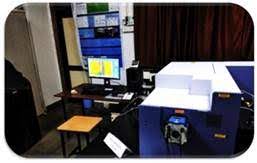
To make the model truly functional, it had to do more than just sit still. In the body, lungs are constantly in motion. AlveoliX developed specialized machinery to subject the chips to rhythmic, three-dimensional stretching.
“This mechanical movement is crucial,” explains Jakson Luk, Postdoctoral Fellow at the Crick Institute and the study’s first author. “It stimulates the formation of microvilli—tiny finger-like projections that increase the surface area of the cells, allowing them to function like real lung tissue.”
Once the “breathing” environment was established, the researchers added a third element: macrophages. These are the immune system’s “first responders” that gobble up bacteria. Crucially, these macrophages were also derived from the same donor’s stem cells, creating a completely autologous (self-derived) system.
Watching TB Take Hold
The team used the chip to study Mycobacterium tuberculosis, the bacteria responsible for TB. Because TB is a slow-progressing disease, the very early stages of infection in humans are nearly impossible to observe in real-time.
Inside the chip, the researchers witnessed a grim architectural shift:
-
Clustering: Macrophages swarmed the bacteria, forming tight clusters.
-
Necrosis: Within five days, the centers of these clusters began to die (necrotic cores), a hallmark of TB progression.
-
System Failure: Shortly after, the barriers between the air sacs and blood vessels collapsed, mimicking the lung failure seen in severe clinical cases.
“We were successfully able to mimic these initial events in TB progression,” says Luk. “It gives us a holistic picture of how different lung cells respond to infections during those unseen early stages.”
Beyond the Lab: Why This Matters for Patients
The implications for public health and drug development are significant. Currently, drug trials rely heavily on animal models, which often fail to predict human responses due to fundamental differences in anatomy and immune function.
1. Personalized “Clinical Trials”
In the future, a doctor could take a blood sample from a patient with a specific genetic profile or a rare lung condition, create a lung-on-chip, and test various antibiotics or antivirals on that specific “person” in a dish before ever prescribing a pill.
2. Replacing Animal Models
“Given the increasing need for non-animal technologies, organ-on-chip approaches are becoming ever more important,” says Max Gutierrez, Principal Group Leader at the Francis Crick Institute. “We can avoid the differences in lung anatomy and immune makeup between animals and humans.”
3. A Blueprint for Other Diseases
While this study focused on TB, the researchers emphasize that the platform can be adapted for:
-
Lung Cancer: Observing how tumors interact with moving lung tissue.
-
COPD and Asthma: Testing how environmental pollutants affect specific genetic groups.
-
Viral Pandemics: Rapidly testing treatments for new respiratory viruses.
Limitations and the Road Ahead
Despite the excitement, experts urge cautious optimism. While the lung-on-chip is a massive leap forward, it does not yet capture the full complexity of the human body.
“A chip lacks a lymphatic system, a nervous system, and the full diversity of the systemic immune response,” notes Dr. Sarah Peterson, a pulmonary specialist not involved in the research. “It is a sophisticated tool for looking at tissue-level interactions, but it doesn’t replace the need for whole-organism study just yet.”
Furthermore, the process of creating iPSC-derived chips is currently expensive and time-consuming, meaning it may be several years before this becomes a standard tool in local hospitals.
The Next Step
The research team is now working on “refining” the chip by adding even more cell types, such as nerve cells and more diverse immune cells, to further close the gap between the plastic chip and the human body.
For the millions living with respiratory conditions, this technology offers a glimpse into a future where “one-size-fits-all” medicine is replaced by treatments as unique as their own DNA.
Medical Disclaimer: This article is for informational purposes only and should not be considered medical advice. Always consult with qualified healthcare professionals before making any health-related decisions or changes to your treatment plan. The information presented here is based on current research and expert opinions, which may evolve as new evidence emerges.
References
Primary Study:
-
Luk, C. H., et al. (2026). “Autologous human iPSC-derived Alveolus-on-Chip reveals early pathological events of M. tuberculosis infection.” Science Advances. DOI: 10.1126/sciadv.aea9874.


