
Bengaluru, India – Researchers at the Indian Institute of Science (IISc) have developed a novel, non-invasive method for estimating glucose concentrations in solutions and tissue using sound waves. This breakthrough, published in the journal Science Advances, offers a potential alternative to the traditional, often painful, needle-prick blood glucose tests that are a daily necessity for millions of people with diabetes.
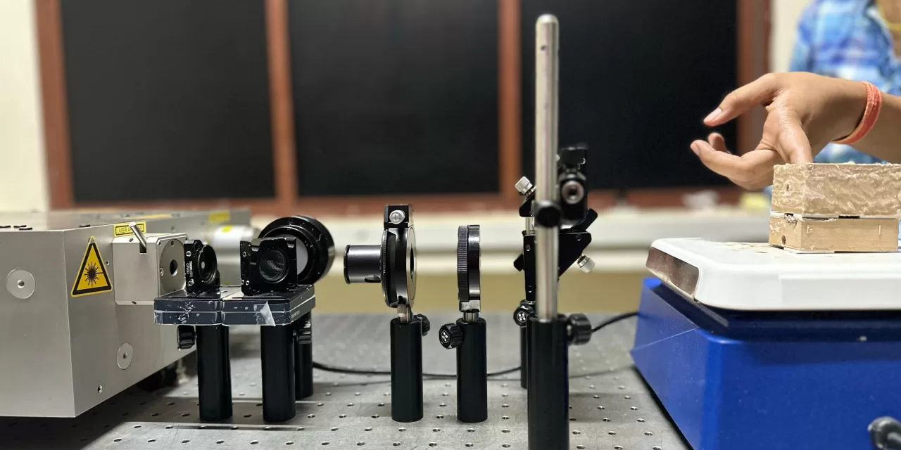
The technique, known as photoacoustic sensing, involves shining a laser beam onto biological tissue. When the tissue absorbs the light, it heats up slightly, causing it to expand and contract, generating ultrasonic sound waves. These sound waves can be detected and analyzed to determine the composition of the tissue.
In this study, the researchers focused on measuring glucose levels using polarized light, a light wave that oscillates in a single direction. Glucose, a chiral molecule with a unique structural asymmetry, rotates polarized light as it passes through it. The team discovered that by changing the orientation of the polarized light, they could observe changes in the intensity of the emitted sound waves.
“We don’t actually know why the acoustic signal changes when we change the polarization state. But we can establish a relationship between the glucose concentration and the intensity of the acoustic signal at a particular wavelength,” explained Jaya Prakash, Assistant Professor at IISc and the study’s corresponding author.
Essentially, the degree to which glucose rotates the polarized light is directly related to its concentration. This rotation is reflected in the acoustic signal intensity, allowing researchers to accurately estimate glucose levels in water, serum solutions, and even animal tissue slices.
The technique demonstrated near-clinical accuracy and the ability to measure glucose concentrations at various depths within tissue, thanks to the minimal scattering of sound waves. A pilot study also showed that the sensor setup could track blood glucose concentrations in a healthy participant before and after meals.
“If we know the speed of sound in this tissue, we can use the time-series data to map our acoustic signals to the depth at which they are coming from,” stated Swathi Padmanabhan, Ph.D. student and first author of the paper.
While the current laser source is bulky and expensive, the researchers are working on developing a more compact and cost-effective version for clinical use. Furthermore, the team believes this technique could be applied to other chiral molecules, including many common drugs, opening up a wide range of applications in healthcare and diagnostics. For example, they demonstrated the ability to estimate the concentration of naproxen, a pain relief medication, in ethanol solution.
Disclaimer: This research is still in its early stages. While the results are promising, further studies and clinical trials are necessary to validate the accuracy and reliability of this technique for widespread use in diabetes management. This technology is not intended to replace current medical diagnostic devices or procedures. Always consult with a healthcare professional for medical advice and treatment.
More information: Swathi Padmanabhan et al, Deep tissue sensing of chiral molecules using polarization-enhanced photoacoustics, Science Advances (2025). DOI: 10.1126/sciadv.ado8012


