

In an extraordinary medical case that has captured national and international attention, doctors at Fortis Memorial Research Institute in Gurugram have successfully performed a complex surgical procedure to remove twin parasitic fetuses—known medically as fetus in fetu—from the abdomen of a one-month-old baby girl. The surgery, completed on July 30, 2025, represents one of the rarest and most challenging cases ever documented globally in pediatric surgery.
The newborn presented with severe malnourishment and dehydration caused by the parasitic fetuses pressing against her vital digestive organs, leading to pain and reduced appetite. The condition, fetus in fetu, is a developmental anomaly where a malformed twin, incomplete and parasitic in nature, grows inside the body of its healthy sibling. Globally, fewer than 300 cases have been reported, typically involving just one parasitic fetus; the presence of two makes this case exceptionally unique and medically significant.
Key Developments and Surgical Challenges
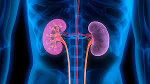
Dr. Anand Sinha, the director of pediatric surgery at Fortis Gurugram and the lead surgeon, described the case as “exceptionally rare and challenging.” “To find not one but two malformed fetuses enclosed within a single sac inside a one-month-old infant was truly exceptional,” he said. The surgery lasted approximately two hours and required meticulous precision to avoid damaging key organs such as the intestines, pancreas, stomach, liver, kidneys, and critical blood vessels.
The infant’s condition had to be stabilized with intravenous nutritional supplements before surgeons could proceed. A multidisciplinary team of about 15 specialists, including pediatric surgeons and anesthesia experts, coordinated to handle this delicate operation. Special precautions, such as constant temperature regulation, were employed because of the baby’s vulnerability, as newborns are particularly sensitive to temperature fluctuations during surgery.
The surgical team successfully extracted the parasitic twins intact, which alleviated the intense pressure on the baby’s stomach and intestines. Dr. Sinha emphasized the use of advanced neonatal surgical techniques and monitoring throughout the procedure to ensure the delicate tissues were preserved. Postoperatively, the baby entered intensive care with close monitoring of vital functions and pain management tailored for newborns.
Medical and Scientific Context
Fetus in fetu is an extremely rare congenital anomaly occurring approximately in 1 out of every 500,000 live births. The condition arises from abnormal embryological development in monochorionic diamniotic twin pregnancies where one twin envelops the other, which then grows in a parasitic fashion inside its sibling. This anomaly must be distinguished from teratomas, which are tumors containing various types of tissues but lack the organization and developmental characteristics seen in fetus in fetu.
The parasitic fetuses in such cases do not have functioning brains, hearts, lungs, or other vital organ systems necessary for independent life. Their growth, however, can cause serious complications for the host twin, including obstruction of vital organs, malnutrition, and life-threatening pressure effects, as seen in this newborn.
Expert Perspectives
International literature reports that surgical excision remains the only definitive treatment for fetus in fetu. Although uncommon, if left untreated, the mass effect caused by the parasitic fetuses can worsen, potentially leading to organ damage and other complications. After successful surgery, the risk of recurrence is minimal, as fetus in fetu is not considered a cancerous growth but rather malformed fetal tissue.
Implications for Public Health and Medical Practice
This successful operation underscores the importance of advanced pediatric surgical facilities and specialized neonatal care units in developing countries like India, enabling the management of rare congenital anomalies once considered difficult or impossible to treat. It also adds to the growing global repository of knowledge, aiding physicians worldwide in the recognition and management of fetus in fetu.
For parents and caregivers, this case emphasizes the importance of early medical consultation and thorough imaging studies, such as ultrasound and computed tomography, when unusual abdominal masses or feeding difficulties are noted in newborns. Early diagnosis allows timely intervention and can significantly improve prognosis.
Potential Limitations and Balanced Viewpoints
Although the surgery was successful, fetus in fetu remains an exceptionally rare condition, limiting the availability of large-scale studies on long-term outcomes. While surgical excision often results in good recovery, close long-term follow-up is essential to monitor for complications or rare recurrences.
Some medical experts caution that fetus in fetu is sometimes misdiagnosed as teratomas, which have different treatments and prognoses. Definitive diagnosis relies on detailed pathological examination and imaging studies, necessitating expert multidisciplinary collaboration.
The case also highlights the delicate balance of timing surgery in very young or medically fragile infants, where risks of anesthesia and operative complications must be carefully weighed against the potential harm of delaying treatment.
References


