CHARLOTTESVILLE, VA — Researchers at UVA Health have developed a groundbreaking MRI technique that offers new insights into the role of fat surrounding the heart, potentially revolutionizing the way heart disease is diagnosed and treated.
The innovative method, spearheaded by Dr. Frederick H. Epstein and his team from the University of Virginia’s Department of Biomedical Engineering, enables doctors to assess the composition of adipose tissue—fat—that encircles the heart. This non-invasive technique could help identify individuals at high risk for serious cardiac conditions such as coronary artery disease, atrial fibrillation (irregular heartbeat), and heart failure, even before symptoms arise.
The study, published in Magnetic Resonance in Medicine, highlights how understanding the makeup of this heart-associated fat can provide valuable insights into a patient’s health. Traditionally, the health risks of excess fat around the waist or hips have been well-known, but the fat around the heart, known as epicardial adipose tissue, has often been overlooked.
“Using this new MRI technique, we now for the first time have the ability to know the composition of the fat that accumulates around the heart. This is important because depending on its makeup, this fat can release harmful substances into the heart muscle, contributing to serious heart problems,” said Dr. Amit R. Patel, a cardiologist at UVA Health and co-author of the study.
The heart is naturally surrounded by a layer of fat, which, when healthy, provides vital support to the organ. However, for individuals with obesity or those at risk for heart disease—due to factors like high blood pressure, smoking, poor diet, and diabetes—this fat can accumulate in excess, become inflamed, and undergo harmful transformations. The UVA team hopes to use MRI to detect and differentiate these fat types, potentially predicting future heart problems.
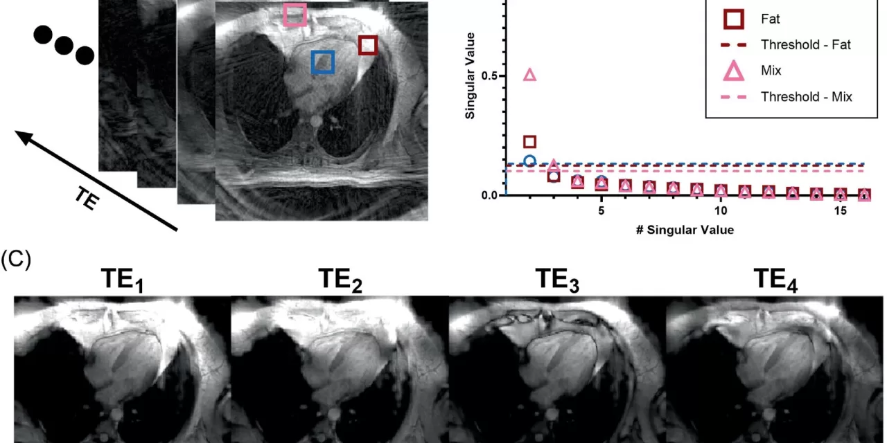
The technique works by analyzing the specific types of fats in the epicardial adipose tissue, such as saturated, monounsaturated, and polyunsaturated fatty acids. This detailed examination could allow doctors to spot at-risk patients long before symptoms of heart disease, the leading cause of death worldwide, begin to manifest.
“This could be a game-changer for diagnosing heart disease,” said Dr. Patel. “By identifying the unhealthy fat surrounding the heart early, we may be able to slow the progression of heart disease through targeted treatments such as diet, exercise, or medication.”
One of the major challenges faced by the research team was the movement of the heart and surrounding organs, which made it difficult to capture clear images of the adipose tissue. However, the researchers overcame this obstacle by developing advanced imaging techniques that capture high-quality scans in just a single breath hold.
“Thanks to the work of biomedical engineering graduate student Jack Echols, we developed computational methods that extract the unique signatures of fatty acids from noisy signals,” Dr. Epstein explained. “This allowed us to see the composition of fat in the heart, which was previously impossible.”
Early tests of the new technique in both the lab and a limited number of human patients have shown promising results. For instance, in patients with obesity and those who had experienced heart attacks, the fat around the heart was found to contain excessive amounts of saturated fatty acids—an indicator of unhealthy fat.
“The ability to visualize the composition of the fat around the heart could be a key tool for identifying at-risk patients and improving our understanding of heart disease,” Dr. Patel added. “Ultimately, this technique could pave the way for new treatment strategies and better outcomes for patients.”
With ongoing research, the UVA team aims to refine the technique and expand its clinical applications, with the hope of improving heart disease management and patient outcomes.
For more information, refer to the study published in Magnetic Resonance in Medicine: John T. Echols et al., Fatty Acid Composition MRI of Epicardial Adipose Tissue: Methods and Detection of Proinflammatory Biomarkers in ST-Segment Elevation Myocardial Infarction Patients (2024). DOI: 10.1002/mrm.30285.


