
January 7, 2026
GURUGRAM – In a medical milestone for Southeast Asia, surgeons at the Fortis Memorial Research Institute (FMRI) have successfully performed a rare, life-saving hybrid cardiac procedure on a 29-year-old man. The complex surgery, which combined traditional open-heart bypass techniques with minimally invasive endovascular repair, saved the patient from a ruptured thoracic abdominal aorta—a condition that carries a near-certain mortality rate if left untreated.
The patient, a resident of Bihar whose identity has been withheld for privacy, arrived at the Delhi-NCR facility in critical condition following months of misdiagnosis and failed interventions at multiple medical centers across India. His recovery and discharge within just six days of the procedure signal a significant shift in how complex vascular emergencies may be managed in the region moving forward.
A Race Against Time: The Patient’s Journey
The case began as a medical mystery in Bihar, where the patient initially presented with severe chest complications. Early assessments misidentified his condition as a simple accumulation of fluid in the chest (pleural effusion). This led to the insertion of a chest tube—a standard procedure for fluid drainage that, in this specific case, exacerbated internal bleeding because the underlying issue was actually a ruptured artery.
By the time the patient reached Gurugram, his heart function had plummeted to a staggering 15%, leaving him bedridden and at constant risk of a “blowout” hemorrhage.
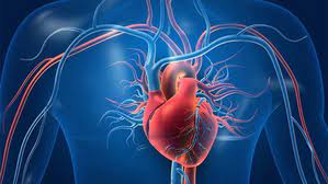
“The thoracic abdominal aorta is the body’s ‘superhighway’ for blood,” explains Dr. Udgeath Dhir, Principal Director of Cardio Thoracic Vascular Surgery at FMRI, who led the surgical team. “When this vessel ruptures, the internal bleeding is usually fatal within minutes. This patient was essentially living on borrowed time.”
Understanding the “Hybrid” Innovation
The surgery was necessitated by the location and severity of the rupture. A thoracic abdominal aortic aneurysm (TAAA) involves the section of the aorta that spans both the chest and the abdomen. It is particularly dangerous because this segment of the artery supplies blood to the kidneys, liver, and intestines.
The Problem with Conventional Surgery
In a standard open repair for such a massive rupture, surgeons must “clamp” the aorta. This stops blood flow to vital organs for an extended period, which carries a 50% risk of:
-
Permanent kidney failure
-
Paralysis (due to lack of blood to the spinal cord)
-
Multisystem organ failure
The Hybrid Solution
To mitigate these risks, the team utilized a Hybrid Approach. This involves two distinct phases performed in a single surgical session:
-
The Open Bypass: Surgeons first performed a bypass to create “detours” for the blood, ensuring that the liver, kidneys, and intestines continued to receive oxygenated blood even while the main artery was being repaired.
-
Endovascular Stent Grafting: Instead of cutting open the entire length of the aorta, surgeons used a minimally invasive technique. They inserted a catheter through the groin to deploy a fabric-covered metal mesh (stent) to seal the rupture from the inside.
Expert Commentary: Why This Matters
While hybrid suites are becoming more common in Western medicine, their application for a ruptured thoracic abdominal aorta is rare in Southeast Asia due to the technical expertise and infrastructure required.
“The success of this case is a testament to the evolution of ‘Total Aortic Care’ in India,” says Dr. Arpan Seth, a vascular specialist not involved in the case. “Managing a patient with 15% heart function is a nightmare for any anesthesiologist and surgeon. Using the hybrid method reduced the ‘cross-clamp’ time, which is likely why the patient avoided paralysis and organ failure.”
However, Dr. Seth notes that while this is a victory, the complexity of such surgeries means they are currently limited to high-volume “Centers of Excellence.”
Public Health Implications and Future Outlook
This case highlights a critical gap in early vascular diagnosis in rural India. The patient was turned away by hospitals in Bihar, Kolkata, and Bengaluru, as the risk of “death on the table” was deemed too high.
Key Statistics
-
Aortic Rupture Mortality: Without surgery, the mortality rate for a ruptured thoracic aorta exceeds 90%.
-
Surgical Risk: Even with modern techniques, the estimated mortality risk for this specific patient was nearly 50% due to his weakened heart.
-
Regional First: This marks the first documented case of this specific hybrid configuration for a ruptured aorta in Southeast Asia.
The patient is currently stable and undergoing medical management to strengthen his heart function. Doctors plan for a secondary cardiac procedure once his body has fully recovered from the trauma of the rupture.
Lessons for the Health-Conscious Consumer
For the general public, this case serves as a reminder of the importance of seeking specialized care for persistent, unexplained chest or abdominal pain.
“Patients shouldn’t be afraid to ask for a second opinion if a treatment isn’t working,” says Dr. Dhir. “What looked like fluid in the lungs was actually a life-threatening arterial break. Advanced imaging like a CT Angiogram can be the difference between a misdiagnosis and a life-saving intervention.”
Reference Section
- https://www.theweek.in/wire-updates/national/2026/01/06/southeast-asias-first-hybrid-cardiac-surgery-saves-29-year-old-in-delhi-ncr-hopsital.amp.html
Medical Disclaimer: This article is for informational purposes only and should not be considered medical advice. Always consult with qualified healthcare professionals before making any health-related decisions or changes to your treatment plan. The information presented here is based on current research and expert opinions, which may evolve as new evidence emerges.


