
Tokyo, Japan – A team of Japanese researchers has made a groundbreaking advancement in medical imaging by developing a millimeter-wave sensor (MWS) capable of non-invasively visualizing respiratory movement during diagnostic X-ray and computed tomography (CT) examinations. This innovative technology could significantly improve image quality and treatment accuracy in radiation therapy and diagnostic imaging.
Monitoring respiratory motion is critical during diagnostic imaging and radiation therapy to ensure accurate diagnosis and precise dose delivery while minimizing exposure to organs at risk. However, the absence of practical, non-invasive tools has traditionally made real-time respiratory monitoring challenging, leading to potential image quality issues and repeat scans.
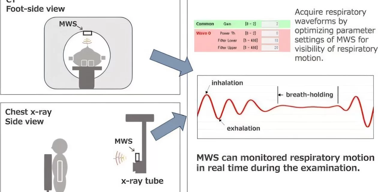
To address this challenge, researchers led by Dr. Hiroyuki Kosaka, alongside Dr. Kenji Matsumoto and Dr. Hajime Monzen from the Department of Medical Physics at Kindai University, developed the MWS. Unlike traditional infrared sensors that require reflective markers on a patient’s body, the MWS is a non-contact device that uses electromagnetic radiation to detect respiratory motion, ensuring patient privacy and comfort.
The research findings were published online in Medical Physics on January 27, 2025.
Validation and Key Features
The research team tested the system using a 24 GHz microMWS and a controlled respiratory motion phantom (QUASAR). The results demonstrated that the MWS could reliably detect respiratory cycles by capturing motion patterns with high accuracy. Further validation was conducted through trials with 20 healthy volunteers aged between 6 months and 64 years, confirming the system’s effectiveness in real-world conditions.
Key advantages of the MWS system include:
- Non-contact monitoring that enhances patient privacy and comfort
- Accurate detection of respiratory motion even through clothing
- Stable measurements in both supine and standing positions
- Cost-effective implementation compared to traditional methods
- Seamless integration with existing X-ray and CT equipment
“This technology has the potential to standardize respiratory monitoring across diagnostic imaging,” said Professor Monzen. “By providing real-time feedback, we can significantly reduce the need for repeat imaging and improve diagnostic accuracy.”
Future Applications and Potential Impact
To further test the MWS’s effectiveness, the researchers utilized a radio-wave dark-box system, which evaluated the sensor’s directionality and precision in detecting movement from various angles. Additionally, they employed the fast Fourier transform technique to isolate and analyze specific breathing signals. By comparing the MWS-detected breathing patterns with QUASAR’s simulated waveforms, the study confirmed the system’s reliability in tracking respiratory motion.
Looking ahead, the MWS system could become a standard tool in hospitals and clinics worldwide, enhancing diagnostic imaging and treatment accuracy. Its low cost, ease of use, and high precision position it as a transformative advancement in medical technology. Additionally, this technology could benefit various patient populations, including children, the elderly, and individuals unable to comply with traditional breath-holding techniques.
“By offering a precise, non-invasive, and cost-effective way to monitor respiratory movements, the MWS can enhance diagnostic accuracy, improve treatment outcomes, and contribute to more efficient health care,” concluded Dr. Kosaka. “This breakthrough has the potential to revolutionize how health care providers approach respiratory motion monitoring, improving both patient experiences and outcomes.”
Disclaimer: This article is based on published research findings and is for informational purposes only. The described technology is still under development and may require further validation before widespread clinical adoption.


