
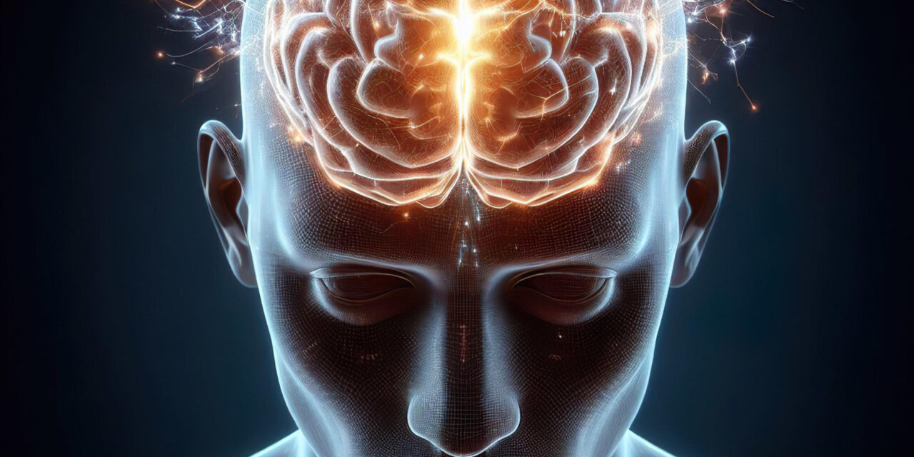
New research challenges longstanding beliefs about how the brain adapts after limb loss, revealing that the brain’s internal map of the body remains stable even after amputation. This discovery offers fresh hope for improving treatments of phantom limb pain and advancing neural-controlled prosthetic limbs.
Who, What, When, Where, Why:
A joint scientific team from the University of Cambridge, UK, and the University of Pittsburgh, US, led by Professor Tamar Makin and Dr. Hunter Schone, has demonstrated that the somatosensory cortex—the brain region responsible for mapping sensory input and body position—retains a virtually unchanged representation of a missing hand even months and years after amputation. The research was published in Nature Neuroscience on August 21, 2025, overturning the previous view that neighboring brain regions reorganize to take over areas representing the lost limb.
Key Findings:
For decades, neuroscience held that after amputation, brain maps reorganize, with adjacent areas expanding into the territory of the missing limb. Evidence behind this came primarily from post-amputation studies without measuring brain maps beforehand, leading to a paradox: many amputees continue to experience phantom limb sensations, including pain, as if the limb still existed. Functional MRI (fMRI) brain scans on three individuals undergoing hand amputation—conducted both before and repeatedly months after their surgery—showed astonishing consistency in the finger and lip maps. Despite the physical loss, the corresponding brain regions still activated almost identically to their pre-amputation state.
The team also studied 26 long-term upper limb amputees, with an average amputation duration of 23.5 years. These individuals displayed brain maps for hand and lips similar to those who had recently undergone amputation, indicating long-term stability of these neural representations.
Expert Perspectives:
Professor Tamar Makin commented, “It seems astonishing that the somatosensory cortex doesn’t ‘know’ the hand is no longer there, maintaining the map in such detail. This challenges the classical belief in brain plasticity following amputation.”
Dr. Hunter Schone noted, “We found no signs of the expected reorganization, with brain maps remaining static. This stability has important implications for treatments of phantom limb pain and prosthetic development.”
Context and Background:
The somatosensory cortex is known to have a body map, a neural representation where different parts of the brain correspond to different parts of the body. However, the study authors caution that these maps have blurry boundaries; multiple neighboring body parts activate overlapping brain regions. Previous research using “winner-takes-all” methods misinterpreted activity from adjacent body parts as brain remapping, explaining the earlier misconception.
Implications for Public Health:
Phantom limb pain—a common and debilitating condition experienced by amputees—has been treated with therapies that attempt to restore or reorganize the brain’s representation of the lost limb. However, clinical trials have shown limited success. The new findings suggest these approaches target the wrong mechanism, as the brain’s map remains intact. Instead, focus may need to shift toward surgical methods that address peripheral nerve health. For instance, grafting nerves to new muscle or skin “homes” to prevent nerve damage and debilitating pain shows promise. One participant who underwent such nerve grafting has since become pain-free.
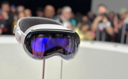
Furthermore, this research bolsters the potential for advanced brain-computer interfaces that harness these stable brain maps to control robotic prosthetics with greater ease and functionality. Dr. Chris Baker from the National Institutes of Mental Health emphasized, “If the brain rewired itself after amputation, these technologies would fail. The stability of body maps provides a real opportunity to develop neural-controlled prosthetics.”
Practical Implications for Readers:
For those living with limb loss or caring for amputees, this study offers new hope for better pain management and improved prosthetic integration in the future. It also highlights the importance of continued innovation in surgical and neural-interface therapies grounded in a refined understanding of brain stability post-amputation.
Limitations and Counterarguments:
While this study represents a significant advance, it involved a small sample size for longitudinal imaging (three individuals) and focused mainly on upper limb amputation. More research is needed with larger and more diverse cohorts to confirm universality. Other brain regions involved in motor control or emotional processing might also undergo changes not captured here. Additionally, the subjective experience of phantom limb pain is complex and may not be fully explained by cortical maps alone.
Medical Disclaimer:
This article is for informational purposes only and should not be considered medical advice. Always consult with qualified healthcare professionals before making any health-related decisions or changes to your treatment plan. The information presented here is based on current research and expert opinions, which may evolve as new evidence emerges.
References:
-
Schone, HR et al. Stable Cortical Body Maps Before and After Arm Amputation. Nature Neuroscience, August 2025. DOI: 10.1038/s41593-025-02037-7
-
Interview with Professor Tamar Makin, Medical Research Council Cognition and Brain Science Unit, University of Cambridge
-
Interview with Dr. Hunter Schone, Department of Physical Medicine and Rehabilitation, University of Pittsburgh
-
Statement from Dr. Chris Baker, Laboratory of Brain & Cognition, National Institutes of Mental Health
This study fundamentally reshapes our understanding of brain plasticity after limb loss and opens new pathways for treating amputees more effectively while leveraging the brain’s remarkable yet stable body maps for technological advancements.


