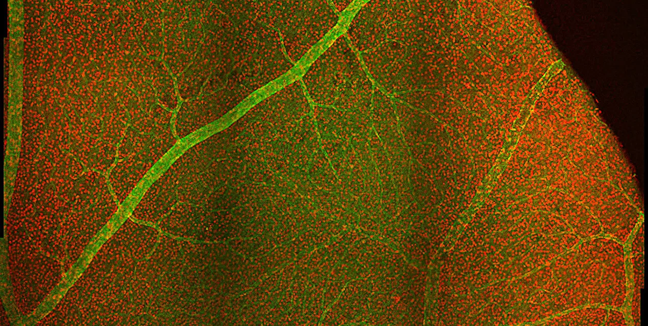
Göttingen, Germany – A groundbreaking study by scientists at the University Medical Center Göttingen (UMG) has revealed that groups of nerve cells in the retina work together to process high-contrast images and movement, challenging the long-standing “efficient coding hypothesis.” Published in Nature, the study could pave the way for novel blindness therapies.
The Retina’s Energy Dilemma
The retina, where light-sensitive photoreceptors and initial nerve cells are located, performs one of the most energy-demanding tasks in the body: processing optical information. For decades, the efficient coding hypothesis posited that the retina minimizes energy use by activating as few nerve cells as possible to relay visual signals to the brain.
However, the research team led by Prof. Dr. Tim Gollisch discovered that this principle does not apply universally. Instead, their experiments showed that specific groups of retinal nerve cells often fire together in response to particular visual stimuli, such as high-contrast images or directional movements.
Redundant Yet Critical Cooperation
The study found that this simultaneous activity, while seemingly inefficient, plays a crucial role in helping the brain distinguish essential visual cues, such as contrast and motion, from less relevant changes like fluctuations in brightness caused by environmental factors.
“This coordinated cooperation of nerve cells allows the brain to prioritize important visual signals over minor distractions,” explained Prof. Gollisch. “At the same time, these groups of cells maintain energy efficiency by reacting only briefly to relevant stimuli.”
Implications for Blindness Therapies
The findings hold promise for developing treatments for blindness, particularly conditions caused by the degeneration of photoreceptors. When photoreceptors die, the nerve cells lose the ability to transmit signals. By artificially activating these cells—such as through visual prostheses or optogenetic therapies—it may be possible to restore vision by mimicking the coordinated activity patterns identified in the study.
“If we can artificially induce the right nerve cell activity, the brain might interpret these signals as naturally as possible, improving outcomes for patients,” said Dr. Dimokratis Karamanlis, the study’s first author and former postdoctoral researcher at UMG.
Testing with Natural Stimuli
The researchers tested retinal tissue samples by projecting natural photographs and simulating eye movements. Measuring the electrical activity of multiple nerve cells, they found that while some cell classes adhered to the efficient coding hypothesis, others consistently acted in unison, defying the principle.
This discovery suggests that the retina employs a dual strategy: energy-efficient coding for less dynamic scenes and coordinated activity for critical stimuli.
Future Applications
The study’s insights are already shaping therapeutic approaches at the Else Kröner Fresenius Center for Optogenetic Therapies in Göttingen. The team aims to use light-sensitive proteins to activate nerve cells, generating natural-like activity patterns for visual restoration.
“These findings will guide us in designing therapies that replicate natural retinal signals, enabling patients to perceive the world more accurately,” said Prof. Gollisch. Clinical trials with patients are expected to begin within a few years.
Reference
Dimokratis Karamanlis et al, Nonlinear receptive fields evoke redundant retinal coding of natural scenes, Nature (2024). DOI: 10.1038/s41586-024-08212-3
This research challenges long-held assumptions and opens new doors in vision science, demonstrating how the eye’s nerve cells adapt to prioritize and convey vital visual information.


