
The tails were a clue. As certain types of mice age, their tails can become stiff and kinked. However, in the lab of molecular biologist Shin-Ichiro Imai at Washington University School of Medicine in St. Louis, the aged mice displayed tails that were flexible and nearly straight. These genetically modified mice seemed to resist aging in other ways too—they were more robust, spent more time running on exercise wheels, and most strikingly, lived about 7% longer than their normal counterparts, gaining an extra 58 days of life. This was reported by Imai and his team earlier this year in Cell Metabolism.
The genetic modification made by the researchers enhanced a crucial communication signal from the brain to the body. This tweak kept a specific group of neurons in the hypothalamus, a key physiological control center deep in the brain, active as the animals aged. Imai’s team discovered that these neurons send signals to the animal’s fat stores via the sympathetic nervous system, a network of nerves that carries messages from the brain throughout the body. In response to this signal, the fat burns lipids and secretes a long-distance signal known as NAMPT, which prevents aging-related damage in other parts of the body, including the hypothalamus itself.
Normally, these neurons lose their signaling ability as mice age, leading to less stimulation of the fat stores. This results in fat accumulation, decreased activity, and reduced NAMPT production, promoting physical decline. Counteracting this communication breakdown appeared to keep the mice more vigorous.
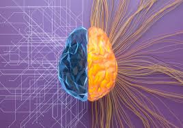
Imai and his team are part of a growing number of scientists studying how communication among organs and tissues, including the brain, liver, muscle, and bone, might combat aging. “We need to understand interorgan communication if we want to understand the aging process,” says geneticist Norbert Perrimon of Harvard Medical School. Last year, the National Institute on Aging requested grant proposals to investigate these connections. Although this research is still in its early stages, there is increasing confidence that faulty communication among organs contributes to aging, says neuroendocrinologist Dongsheng Cai of the Albert Einstein College of Medicine.
Several labs have started testing ways to fix interorgan communication problems in animals. If these findings can be applied to human aging, researchers hope similar interventions could slow our own physical deterioration.
Over the past couple of decades, researchers have found that long-distance signaling involves more than just a few glands and organs like the pancreas, thyroid, testes, and ovaries, which release familiar hormones such as insulin and testosterone. Most organs and tissues now seem to send each other a vast array of molecular messages. For example, fat sends out more than 100 different molecules, and muscles send out over 600. Even bones, once thought to be silent, communicate with muscles, the brain, and other organs.
The nervous system is also part of this expanding signaling network. Researchers have identified numerous new molecular signals and revised centuries-old maps of the nervous system to include newly discovered connections. “We are redefining anatomy,” says neuroscientist Ana Domingos of the University of Oxford.
This organ-to-organ communication can significantly impact health. For instance, when patients develop pancreatitis, an inflammation of the pancreas, there’s a 50% chance that the inflammation will spread to the lungs, making pancreatitis fatal in half of these cases, says biochemist and molecular biologist Stefan Rose-John of the Christian-Albrecht University of Kiel.
Scientists have only begun to catalog the body’s array of signals. Blood, for example, is rich with molecules that may act as interorgan messages, influencing physiology or aging. Even substances that don’t resemble traditional signals might serve as messengers, as seen when gases like nitric oxide were found to affect organs. However, merely identifying signaling molecules in the blood isn’t enough. Molecular biologist Ilia Droujinine of Scripps Research says, “If you take a snapshot of the blood, you don’t know where the proteins are coming from and where they are going.”
In 2021, Droujinine, Perrimon, and colleagues unveiled a technique to identify potential interorgan signals. They genetically modified a particular organ in an animal so that its cells attached a marker, the molecule biotin, to proteins destined to be secreted. These tagged proteins carried a badge of their origin as they traveled through the body. Testing this technique in fruit flies, the researchers found that 51 proteins moved from the muscles to the head, and 269 moved from the fat body, the insects’ main energy storehouse, to the legs. A follow-up 2022 study showed similar results in mice.
Not all proteins identified by this technique are necessarily signals, Droujinine cautions. Some may just be floating in the blood. But once a comprehensive list of interorgan signals is compiled, researchers can systematically investigate how these signals change with aging.
Working with fruit flies, molecular biologist Hongjie Li of Baylor College of Medicine and colleagues have developed another method to build this list. They sequence the messenger RNA (mRNA) molecules from all cells in a fly’s body. By analyzing the types of mRNA, the researchers can identify signal molecules and their receptors, map them to specific organs, and infer potential communication routes. For instance, if a fly’s muscles produce a particular protein signal and its fat has the corresponding receptor, the two may be communicating.
In a recent preprint posted to bioRxiv, Li and his team identified three of the brain’s communication partners in the insects. Previous studies had already identified two of them, the fat and the gut, but Li’s group discovered that the brain also communicates with the reproductive organs. “After all, most of our body parts are more or less controlled by the brain,” Li says. The researchers established the existence of this communication channel with multiple lines of evidence.
A clearer picture of brain communication with distant organs is also emerging. Early anatomists mapped the nervous system through dissection, Domingos says. However, better microscopes, improved markers for labeling neuron types, and new techniques to make an animal’s body transparent have revealed new circuits and ruled out previously assumed connections.
For example, evidence suggested that sympathetic nerves connect to white adipose tissue, the main type of fat, but researchers couldn’t confirm this in living animals. In 2015, Domingos and colleagues observed living mice and found that nerve fibers directly connect to fat cells.
Domingos’s team also identified a message conveyed by these connections. When sympathetic neurons were stimulated, fat cells responded by breaking down lipids. Previously, researchers thought hormones stimulated this process. But Domingos’s study and other findings, including Imai’s, indicate that the sympathetic nervous system plays a larger role in metabolism than expected.
This brain-to-body link weakens with age, Imai’s team showed. As mice age and neurons in the hypothalamus become less active, sympathetic nerves retract from white adipose tissue, reducing nerve stimulation and NAMPT production, potentially creating a cycle of dysfunction. The cause of this decline is unclear, but it might be due to age-related inflammation in the hypothalamus, white adipose tissue, or both.
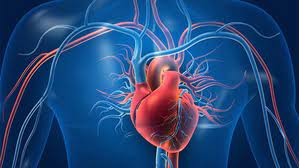
The heart also loses key nerve connections with the brain as mice age, cardiovascular biologist Stefanie Dimmeler of Goethe University Frankfurt and her team revealed in 2023. They found that as mice aged, nerve fibers disappeared from the left ventricle, the chamber that pumps blood to most of the body. Instead of dying, the fibers retracted, uncoupling from the heart muscle, reducing heart rate variability. This decline also occurs in humans and is linked to early death.
Senescent cells, damaged cells that accumulate with age, trigger the nerves’ retreat, Dimmeler and colleagues found. Eliminating these cells in mice’s hearts with two drugs reconnected the nerves. Determining if human hearts experience a similar nerve loss is challenging due to the difficulty in obtaining healthy human hearts, Dimmeler says.
Researchers have found other instances where altered interorgan communication in lab animals promotes physical decline or accelerates aging. Geneticist Hua Bai of Iowa State University and colleagues discovered that as fruit flies age, their equivalent of the liver releases more inflammation-promoting cytokines. This protein causes the insects’ hearts to beat abnormally. In mammals, the equivalent cytokine IL-6 increases with age, suggesting a similar effect could occur in humans.
Another lab favorite, the nematode Caenorhabditis elegans, provided more evidence linking aging with organ communication. Mitochondria, the cell’s energy producers, can suffer damage from reactive oxygen species and other causes. Damaged proteins can disrupt energy production and spark the mitochondrial unfolded protein response to restore proper folding and destroy damaged proteins.
Cells also send out warning messages that trigger the unfolded protein response in mitochondria of distant tissues, molecular biologist Andrew Dillin of the University of California, Berkeley, and colleagues found. This protective effect boosts longevity. Last year, Dillin and colleagues revealed that stimulating the unfolded protein response in certain nervous system cells also occurred in the intestines, extending some worms’ lives by more than 20%.
However, evidence suggests the mitochondrial unfolded protein response can become too effective with age and destructive.
If these communication channels are as significant in humans as in animals, they could inspire new ways to treat diseases and possibly counteract aging. Li envisions treatments for neurodegeneration based on his fruit fly studies. Potential therapies often can’t cross the blood-brain barrier, but treating an easier-to-access organ could pass benefits to the brain via messages that can cross the barrier.
Of the thousands of interorgan messages in the body, a few show promise for therapies. Osteocalcin, a hormone produced by bones, might counteract aging effects. Initially expected to strengthen bones, osteocalcin was found to induce various metabolic benefits, including stimulating insulin release and glucose absorption by muscles during exercise. Mice lacking osteocalcin failed memory tests, suggesting it is crucial for cognition.
“Osteocalcin could counteract some manifestations of aging,” Karsenty says. He and colleagues are studying mice


