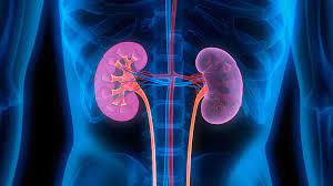
Pohang, South Korea – A research team at Pohang University of Science and Technology (POSTECH) has achieved a groundbreaking advancement in medical imaging by employing ultrafast ultrasound, capturing 1,000 images in just one second. The team, led by Professor Chulhong Kim, has successfully unraveled the mysteries of kidney diseases using this cutting-edge technology, capable of visualizing the three-dimensional microvasculature of the kidneys without the need for contrast agents.
The kidneys, crucial for filtering waste and maintaining the balance of substances in the bloodstream, are susceptible to conditions like hypertension and diabetes. Compromised kidney function can lead to irreversible kidney failure, necessitating lifelong treatments such as artificial hemodialysis or kidney transplantation. The research team’s novel technique, which offers a non-invasive and contrast-agent-free method of imaging, could play a pivotal role in preventing and managing kidney failure.
Conventional medical imaging methods like CT (computed tomography) and MRI (magnetic resonance imaging) have limitations in capturing fine vascular structures due to resolution and sensitivity constraints. Additionally, the use of contrast agents in these methods poses risks for patients with kidney disease. In contrast, ultrasound imaging, known for its safety in fetal monitoring, measures real-time blood flow velocity and direction without the need for contrast agents.
However, the speed of current ultrasound imaging has limitations in capturing fine blood vessels with sufficient sensitivity. The POSTECH research team addressed this challenge by enhancing microvascular sensitivity with ultrafast ultrasound acquisition, capturing an impressive 1,000 frames per second, over 100 times faster than conventional ultrasound imaging.
In a significant achievement, the researchers successfully imaged the entire three-dimensional vascular network of the renal artery, vein, and interlobular arteries and veins in the renal cortex, all without the use of contrast agents. The technique allowed continuous observation of renal vascular changes in an animal model with induced renal failure.
Professor Chulhong Kim highlighted the potential applications of this system, stating, “The system allows us to understand the pathophysiology of diseases leading to kidney failure, enabling the observation of vascular changes before and after kidney transplantation.” He emphasized its broader implications, noting its potential use in studying blood circulation and functional impairment across various organs, including the digestive system, circulatory system, and cerebral nervous system.
This research was supported by grants from the National Research Foundation (NRF), the Korea Medical Device Development Fund, the Korean Fund for Regenerative Medicine, and BK21 FOUR projects funded by the Korean government.


