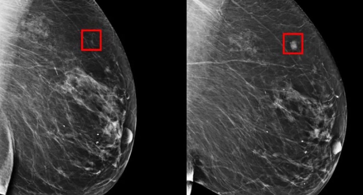
Atlanta, GA – A groundbreaking study presented at the American College of Cardiology’s Annual Scientific Session (ACC.25) reveals that mammograms, aided by artificial intelligence (AI), can provide valuable information beyond breast cancer detection, offering a new window into cardiovascular health.
Researchers have demonstrated that AI models can analyze mammogram images to assess the amount of calcium buildup in breast arterial tissue, a significant indicator of cardiovascular risk. This innovative approach leverages the existing infrastructure of routine mammogram screenings, potentially transforming them into dual-purpose tools.
“We see an opportunity for women to get screened for cancer and also additionally get a cardiovascular screen from their mammograms,” said Theo Dapamede, MD, PhD, a postdoctoral fellow at Emory University and the study’s lead author.
The U.S. Centers for Disease Control and Prevention recommends regular mammograms for middle-aged and older women, resulting in approximately 40 million screenings annually in the U.S. While breast arterial calcifications are visible in these images, they are not routinely quantified or reported. This study introduces an AI-powered image analysis technique that can automatically analyze these calcifications and generate a cardiovascular risk score.
The AI model was trained using a large dataset of over 56,000 mammogram images and electronic health records from Emory Healthcare, spanning 2013 to 2020. This extensive dataset allowed the model to accurately segment calcified vessels and predict the risk of cardiovascular events.
The study found that the AI model effectively categorized patients’ cardiovascular risk as low, moderate, or severe based on mammogram images. Notably, the model showed that women with the highest level of breast arterial calcification had a significantly lower five-year event-free survival rate compared to those with the lowest level.
“Our study showed that breast arterial calcification is a good predictor for cardiovascular disease, especially in patients younger than age 60,” Dapamede stated. “If we are able to screen and identify these patients early, we can refer them to a cardiologist for further risk assessment.”
The researchers believe this tool could be particularly beneficial for younger women, who can benefit most from early interventions. Future research will explore the potential of similar AI models for assessing biomarkers related to other conditions, such as peripheral artery disease and kidney disease, from mammogram images.
The AI model was developed through a collaboration between Emory Healthcare and Mayo Clinic. It is not currently available for public use. If it successfully passes external validation and secures approval from the U.S. Food and Drug Administration, the tool could be made commercially available for integration into routine mammogram processing.
Disclaimer:
It is important to note that the AI model discussed in this article is currently under development and is not yet available for clinical use. The information presented is based on preliminary research findings and should not be interpreted as medical advice. The availability and efficacy of this technology are subject to further validation and regulatory approval. Individuals with concerns about their cardiovascular health should consult with a qualified healthcare professional.


