
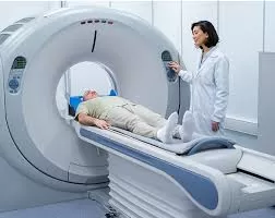
Magnetic resonance imaging (MRI) is an indispensable tool in modern medicine, providing clinicians with detailed insights into the human body and aiding in the diagnosis of various conditions. However, differences in imaging protocols and equipment across medical institutions present significant challenges in ensuring consistent and reliable interpretation, particularly in multi-center research studies.
To address this challenge, a groundbreaking study led by Dr. Gregory Lodygensky, a clinical professor at Université de Montreal and clinician-researcher at Sainte-Justine Hospital, has proposed a method to standardize MRI images across different hospitals. Collaborating with professor-researchers Jose Dolz and Christian Desrosiers from the École de technologie supérieure (ETS), their work—published in Medical Image Analysis—introduces an innovative approach to modifying MRI images, making them more comparable across various institutions.
The Challenge of MRI Variability
Each hospital, clinic, or research facility employs unique MRI imaging protocols based on its available equipment and specific scanning parameters. These variations lead to inconsistencies in contrast, brightness, and other image properties, creating significant obstacles when researchers attempt to pool data from multiple sources.
The new harmonization method aims to mitigate these inconsistencies, ensuring that MRI images from different institutions can be analyzed more reliably, thus enhancing the accuracy of medical research and clinical diagnostics.
A Three-Step Approach to Harmonization
The harmonization method, developed by ETS doctoral candidate Farzad Beizaee, follows three key steps:
- Learning the Source Domain: A model is trained to understand how MRI images from a specific machine or institution are structured and distributed.
- Reformatting External MRIs: The model then adjusts MRI images from different institutions to align with the learned distribution while preserving unique patient-specific details.
- Adapting to New Data: When new images from an unfamiliar machine are introduced, the model ensures that they still conform to the original learned distribution.
Testing and Validation
To validate this technique, the researchers tested it using MRI brain scans from U.S. databases and a neonatal imaging consortium in Australia. The model was applied to two specific tasks: segmenting brain images to analyze structural consistency and estimating brain age in newborns. The results demonstrated that the new technique outperformed existing harmonization methods, showing its adaptability across diverse patient groups.
Notably, this approach successfully processed MRI images of newborns with brain lesions—an achievement that existing models have struggled to accomplish due to their training on healthy brain images.
“Thanks to this model, we can now interpret data from thousands of families and children monitored at different hospitals, despite variations in MRI scanners,” said Dr. Lodygensky. “This advancement significantly improves our ability to analyze large cohorts, a challenge that has hindered medical research for years.”
The Future of MRI Harmonization
The success of this method opens doors for future large-scale applications. Dr. Lodygensky and his team plan to extend their research to broader datasets, further refining the model’s ability to enhance medical imaging consistency. Ultimately, this innovation could improve the accuracy of medical diagnoses and research findings worldwide.
Reference
Beizaee, F., et al. (2025). Harmonizing flows: Leveraging normalizing flows for unsupervised and source-free MRI harmonization. Medical Image Analysis. DOI: 10.1016/j.media.2025.103483
Disclaimer: This article is based on a published research study and is intended for informational purposes only. It does not constitute medical advice or professional consultation. Readers are encouraged to refer to the original research and consult healthcare professionals for further information.


