
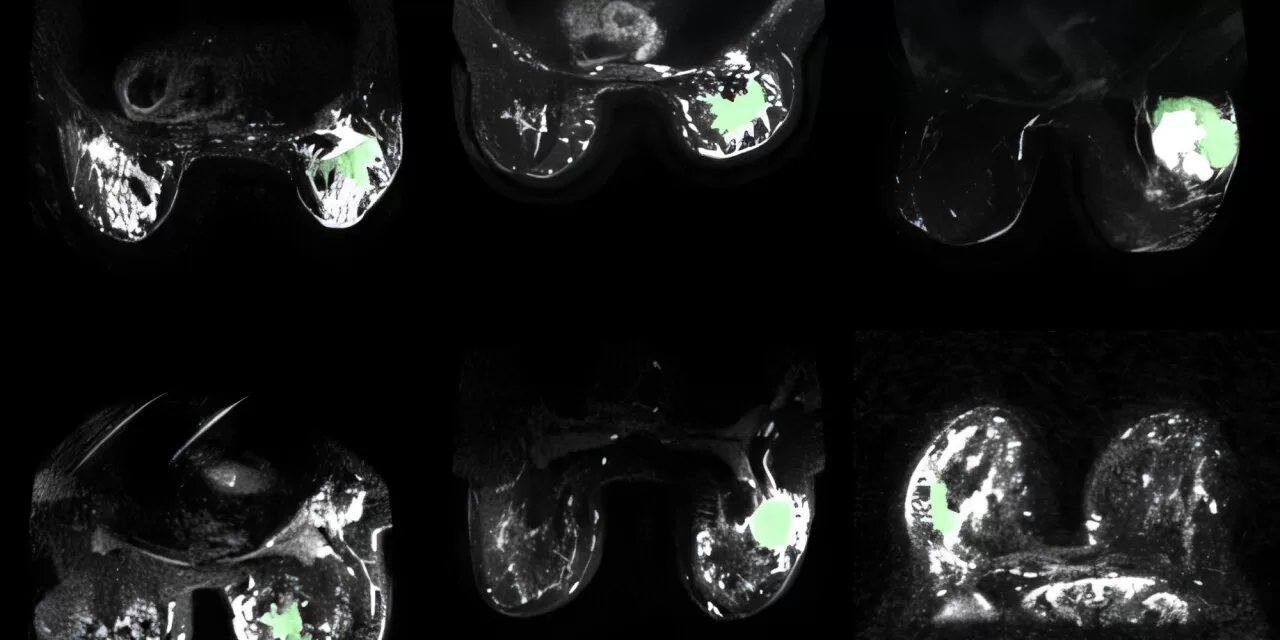
Waterloo, Canada – In a breakthrough for breast cancer detection and treatment, researchers at the University of Waterloo have optimized an artificial intelligence (AI)-enhanced magnetic resonance imaging (MRI) technology that makes cancerous breast tissue glow in scans.
Originally developed for prostate cancer, this innovative imaging method has now been fine-tuned using AI to improve its accuracy in identifying cancerous tissue in the breast. The research, published in the journal Sensors, demonstrates how this advancement can help doctors and radiologists pinpoint tumors with greater precision.
“This technology has great potential to not only improve breast cancer detection, but also treatment,” said Dr. Alexander Wong, a professor in the Department of Systems Design Engineering at the University of Waterloo and co-director of the Vision and Image Processing (VIP) Lab. “Our images contain key predictive information to help clinicians determine the best courses of action for treating each patient.”
How It Works
The system, known as synthetic correlated diffusion imaging (CDI), leverages specific physical characteristics of breast tissue, such as density and the irregular packing of cells. These properties influence the movement of water molecules, which behaves differently in cancerous tissue compared to healthy tissue. CDI enhances these differences by capturing, synthesizing, and mixing MRI signals at various gradient pulse strengths and timings, creating images with clear distinctions between healthy and cancerous tissues.
By offering significantly improved delineation of cancerous tissue, CDI provides a powerful tool for radiologists and surgeons. “The more accurate we make CDI for distinguishing between cancerous and healthy tissue, the more effective patient treatment plans can be,” said Amy Tai, a Waterloo engineering Ph.D. student supervised by Wong.
Enhancing Treatment and Surgical Outcomes
One of the primary advantages of CDI-enhanced imaging is its potential to aid surgeons in determining tumor margins with greater accuracy. By providing clearer images, this technology could help surgeons limit the amount of tissue removed during operations while ensuring complete tumor extraction, reducing the likelihood of requiring additional surgeries.
To refine the system for breast cancer, researchers utilized pre-treatment images from more than 350 patients across 10 medical institutions, as part of a study by the American College of Radiology Imaging Network. With promising results, researchers now aim to extend CDI’s applications to other cancers, including those affecting the brain, neck, and head.
“We have already illustrated great potential for prostate cancer, and now we are seeing promising results for breast cancer,” Wong added. “It’s extremely encouraging that we’ll be able to expand and help in other areas as well.”
Disclaimer
This article is for informational purposes only and does not constitute medical advice. Readers should consult healthcare professionals for diagnosis and treatment options related to breast cancer.


