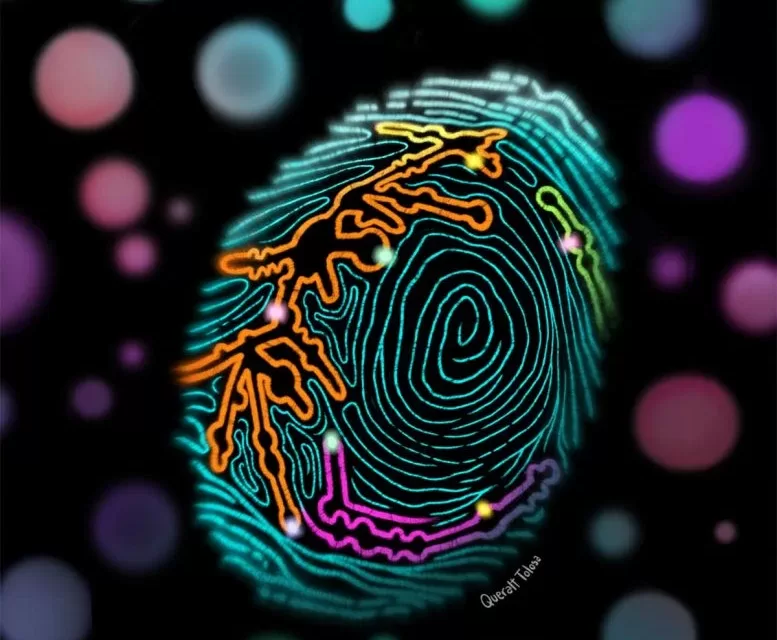
Barcelona, Spain — A groundbreaking study published today in Molecular Cell reveals that scientists have uncovered a cancer “fingerprint,” offering a promising new method for early cancer detection with unparalleled accuracy. This breakthrough, spearheaded by researchers at the Centre for Genomic Regulation (CRG) in Barcelona, could transform how cancer is diagnosed, paving the way for non-invasive tests that can identify various types of cancer earlier than ever before.
The study centers around the ribosome, the protein factories inside cells, which for years were believed to have a uniform structure across the human body. However, researchers discovered a hidden layer of complexity within ribosomes: tiny chemical modifications in ribosomal RNA (rRNA) that differ depending on tissue type, developmental stage, and disease state.
“Our ribosomes are not all the same. They are specialized in different tissues and carry unique signatures that reflect what’s happening inside our bodies,” said Dr. Eva Novoa, ICREA Research Professor at CRG and lead author of the study. “These subtle differences can tell us a lot about health and disease.”
A Hidden Complexity in Cancer Cells
The breakthrough came when scientists analyzed the rRNA from human and mouse tissue samples, including the brain, liver, heart, and testis, uncovering unique patterns of rRNA modifications. These variations, called ‘epitranscriptomic fingerprints,’ act as molecular signatures that can reveal not only the origin of the tissue but also its disease state.
In particular, the team observed these fingerprints in cancer cells, notably lung and testicular cancer, where rRNA modifications were found to be “hypomodified”—meaning cancer cells consistently lose some of these chemical marks.
Dr. Ivan Milenkovic, first author of the study, explained, “The fingerprint on a ribosome tells us where a cell comes from. It’s like each tissue leaves its address on a tag in case its cells end up in the lost and found.”
Early Cancer Detection with Near-Perfect Accuracy
Focusing on lung cancer, the researchers confirmed their findings by comparing the rRNA from healthy and cancerous tissues of 20 patients with stage I or II lung cancer. They found that the rRNA from cancer cells was significantly different, showing promise as a diagnostic biomarker.
Using these molecular fingerprints, the team trained an algorithm capable of accurately distinguishing between cancerous and healthy tissue, achieving near-perfect accuracy in detecting lung cancer at its early stages. “Most lung cancers aren’t diagnosed until later stages, but this test can detect it much earlier, potentially saving valuable time for patients,” said Dr. Milenkovic.
Nanopore Sequencing Technology: A Game Changer
This discovery was made possible by advances in nanopore sequencing technology, a technique that allows researchers to directly analyze rRNA molecules along with their chemical modifications. Unlike traditional methods that removed these modifications, nanopore sequencing enables the real-time capture and study of these molecular changes, providing a more accurate and natural view of the rRNA.
Dr. Novoa explained, “We’ve taken this data out of the junkyard and turned it into a gold mine. It’s an incredible turnaround, especially when information about chemical modifications is captured.”
One of the major advantages of this technology is its portability. Nanopore sequencing devices are small enough to fit in the palm of a hand, making it possible to conduct on-site analysis with minimal samples. In the study, the researchers were able to detect the cancer fingerprint with as few as 250 RNA molecules, a fraction of what typical devices require.
Toward Non-Invasive Diagnostics
Looking to the future, the team hopes to develop a diagnostic test that can detect cancer by analyzing circulating RNA in blood samples, making the process less invasive. Such a test could revolutionize how cancers are detected, requiring only a blood draw instead of tissue biopsies.
However, the authors caution that more research is needed before this approach can be used in clinical settings. “We’re just scratching the surface,” said Dr. Milenkovic. “Larger studies are required to validate these biomarkers across diverse populations and cancer types.”
The Future of Cancer Research
The researchers are also exploring why these rRNA modifications change in cancer cells. Understanding how these changes contribute to the uncontrolled growth and survival of cancer cells could lead to new therapeutic approaches aimed at reversing these harmful modifications.
“We are slowly but surely unraveling this complexity,” said Dr. Novoa. “It’s only a matter of time before we can start understanding the language of the cell.”
This research represents a major step forward in cancer detection and treatment, offering hope for more accurate, earlier diagnoses and paving the way for potentially life-saving advancements in cancer care.
Reference:
“Epitranscriptomic rRNA fingerprinting reveals tissue-of-origin and tumor-specific signatures” by Ivan Milenkovic, Sonia Cruciani, Laia Llovera, et al., Molecular Cell, December 10, 2024.
DOI: 10.1016/j.molcel.2024.11.014


