A groundbreaking artificial intelligence (AI) tool promises to revolutionize maternal and neonatal healthcare by enabling rapid analysis of placentas at birth. Developed by researchers at Northwestern Medicine and Penn State, the tool, PlacentaVision, uses computer vision to detect abnormalities in placental photographs, potentially saving lives through early intervention. The research, published in the journal Patterns, highlights the tool’s potential to improve outcomes for mothers and babies worldwide.
A Faster, Smarter Approach to Placental Analysis
The placenta, a critical organ during pregnancy, is often discarded without examination—especially in resource-limited settings. This oversight misses key opportunities to diagnose infections or conditions like neonatal sepsis, a life-threatening issue affecting millions of newborns globally.
“Placenta is one of the most common specimens in the lab,” said Dr. Jeffery Goldstein, co-author and director of perinatal pathology at Northwestern University Feinberg School of Medicine. “This tool allows us to provide diagnoses days earlier than traditional methods, which can significantly impact medical decisions in urgent cases.”
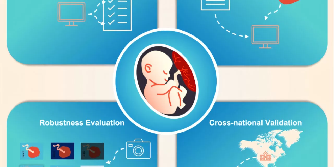
Using a simple photograph, PlacentaVision identifies abnormalities linked to infections or complications. By integrating cross-modal contrastive learning—a method aligning visual images with text data like pathology reports—the AI system delivers high accuracy, even across diverse populations and conditions.
A Solution for Global Health Challenges
The innovation addresses gaps in care for underserved areas. In many regions, women give birth at home without access to medical resources, and placental examinations are rare. Dr. Alison D. Gernand, associate professor at Penn State, emphasized the tool’s potential in such settings.
“Discarding the placenta without examination is a missed opportunity to reduce complications and improve outcomes for mothers and babies,” Gernand said. “This tool could bring advanced diagnostics to areas where they’ve never been possible.”
In addition to aiding low-resource settings, PlacentaVision has applications in well-equipped hospitals, where it can streamline pathology workflows by prioritizing cases needing detailed examination.
Technical Challenges and Innovations
Developing PlacentaVision required addressing significant challenges, such as ensuring the AI model’s adaptability to varying delivery conditions, including poor lighting or low-quality images. Researchers enhanced its robustness using a 12-year dataset of placental images, simulating diverse photo-taking scenarios to refine the tool’s performance.
“The tool needs to be accurate across different environments, from urban hospitals to remote clinics,” explained James Z. Wang, a lead investigator from Penn State. “This flexibility ensures it works in real-world conditions.”
Next Steps: Bringing the Tool to Clinicians
The research team is now working on a user-friendly app for healthcare professionals. Designed to work with minimal training, the app would allow clinicians to photograph a placenta and receive instant feedback. Future enhancements include integrating additional placental features and clinical data to further improve diagnostic accuracy.
“This tool could transform how placentas are examined, especially in parts of the world where such exams are rare,” Gernand said. “With further refinement, PlacentaVision has the potential to save lives and improve long-term health for mothers and infants.”
The tool is poised to bridge critical gaps in maternal and neonatal care, offering a scalable solution for diverse healthcare settings globally.
For more details, refer to the original study: Cross-modal contrastive learning for unified placenta analysis using photographs by Yimu Pan et al., published in Patterns.

