New Method for Liquid Biopsies Offers a Breakthrough in Cancer Detection and Monitoring
October 2024 — In a significant leap forward for cancer diagnostics, scientists at the University of Rochester have unveiled a groundbreaking method that could revolutionize cancer detection and treatment monitoring. By using a single drop of blood, the new technique promises to make cancer testing faster, more affordable, and potentially life-saving.
The method, known as Catch and Display for Liquid Biopsy (CAD-LB), utilizes ultrathin membranes to capture extracellular vesicles (EVs), tiny packets of cellular material released by cells into bodily fluids like blood and saliva. These EVs carry vital information about the body’s health, including proteins and genetic material, which can be key indicators of disease.
For years, scientists have recognized the potential of EVs in medical diagnostics and therapy monitoring. However, previous techniques for isolating and analyzing these vesicles were time-consuming, complex, and costly. The new CAD-LB method addresses these challenges by simplifying the process, enabling quicker, more efficient testing.
Simplified Process for Liquid Biopsies
“By searching blood or other bodily fluids for these extracellular vesicles and the biomarkers they carry, you can find important clues that something is wrong in the body,” said James McGrath, the William R. Kenan Jr. Professor of Biomedical Engineering and leader of the study. “Previously, isolating EVs required many purification steps, making the process slow and complicated. CAD-LB is much simpler and faster, bringing it closer to practical clinical use.”
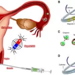
The process is as simple as it is efficient. Blood or bodily fluid samples are quickly prepared, then pipetted onto a special ultrathin membrane. These membranes are designed with perfectly sized pores that catch the EVs. Once the sample is applied, it is analyzed directly under a microscope. By observing which pores glow with biomarkers associated with a particular disease, clinicians can rapidly determine the disease’s prevalence in the body.
A Promising Tool for Cancer Diagnosis
CAD-LB shows great promise for early cancer detection, offering the potential to identify cancers at a curable stage. “This technology is sensitive enough to detect certain cancers early, suggesting it could play a role in cancer screening,” said Jonathan Flax, co-author of the study and research assistant professor at the University of Rochester Medical Center’s Department of Urology.
Beyond detecting cancer, the CAD-LB method could also help tailor personalized treatments for cancer patients. The technique can identify immune modulatory proteins on EVs, which are critical in the body’s ability to fight tumors. These proteins could also predict how a patient might respond to immunotherapies, which are treatments that activate the immune system to target and eliminate cancer cells.
The Road Ahead
The team’s research, published in Small in October 2024, demonstrates the power of CAD-LB to not only diagnose cancer but also to help guide treatment decisions. “It may be used to predict the most effective immunotherapy for a patient, which could greatly enhance the success of their treatment,” said Flax.
As this technology moves toward clinical application, it holds the promise of significantly improving the speed and affordability of cancer detection, providing doctors with powerful new tools to diagnose and treat cancer more effectively than ever before.
For more information, read the full study: “Rapid Assessment of Biomarkers on Single Extracellular Vesicles Using ‘Catch and Display’ on Ultrathin Nanoporous Silicon Nitride Membranes” in Small (DOI: 10.1002/smll.202405505).

